Anterior Teeth
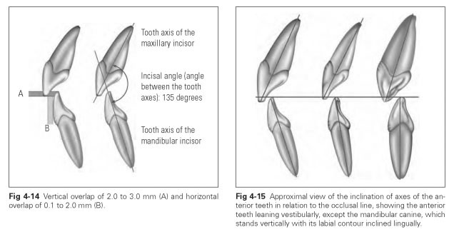
The anterior teeth are the four incisors and two canines in the maxilla and mandible. These 12 teeth form a group of antagonists that produce an interlocking pattern in a normal dentition. When viewed approximally, the maxillary anterior teeth are anterior to and occlude vertically over the mandibular anterior teeth without any contact. The overlap in the horizontal plane is sometimes called the overjet and ranges from 0.1 to 2.0 mm; the vertical overlap is sometimes called the overbite and can be between 2.0 and 3.0 mm (Fig 4-14).
From the approximal view, the interincisal angle is the angle of approximately 135 degrees between the axes of the vestibularly inclined maxillary and mandibular anterior teeth (Figs 4-14 and 4-15). The resulting profile view of the dentition looks strongly like a rudimentary snout. The maxillary canine is inclined so far in a vestibular direction that its tip and the cervical margin lie almost vertically on top of each other and, when viewed vestibularly, give the impression that the labial surface is vertical.
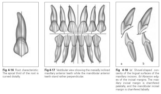
Root characteristic refers to the distal bend of the roots (seen from the vestibular view) (Fig 4-16).The incisors have only one root.The inclinations of the axes of the maxillary anterior teeth seen from the vestibular view show a consistent mesial tendency, while the mandibular anterior teeth stand rather vertically (Fig 4-17). The maxillary canines and central incisors are equally long and the lateral incisors rather shorter; the mandibular anterior teeth lie with their incisal margins on a horizontal line.
Incisors have chisel-, paddle-, or shovel-shaped crowns (Fig 4-18a).The labial and lingual surfaces lean incisally toward each other so that a cutting edge is formed.The approximal surfaces thus appear almost triangular, with the apex of the triangle lying at the incisal margin.
Labial surfaces display the vertical curvature that protects the marginal periodontium, while the lingual surfaces form a tubercle toward the cervix that performs the same function. The incisors are ideal for shearing and cutting through food because of their shovel shape and prominent cutting edge. The incisal margins have an oblique abrasion edge, receding palatally in the maxilla and labially in the mandible so that during anterior movements of the mandible, the incisors slide along on these grinding surfaces (Fig 4-18b).
Mandibular incisors are smaller and narrower than the maxillary incisors; in fact, they are the smallest teeth in the human dentition. The mandibular central incisor, unlike its maxillary counterpart, is smaller than the adjacent lateral incisor. Because they resemble each other very closely, the mandibular central and lateral incisors can be described together. Both teeth have one root, which is significantly flattened in the mesiodis-tal direction and shows a marked longitudinal groove on the outer surface. They are very weak and short and hence unsuitable for post crowns.
Canines are single-rooted teeth that, unlike the incisors, have a cusp tip and are thus dentes cus-pidati. They are the strongest single-rooted teeth. The root of the maxillary canine is the longest in the whole dentition. In rare cases, these teeth show stunting or furcation involvement; very rarely the canine is absent.
The canine is an independent tooth form. No animal with a dentition comprising different tooth forms (heterodont dentition) has more than four canines. When the permanent teeth erupt, the canine often shifts, usually being displaced in a lingual or vestibular direction.
The canines form the corners of the dentition: They have an angled incisal margin or masticatory edge, but at the same time they mark the transition between the incisors and teeth with occlusal surfaces, forming a cornerstone between two very different types of teeth.They also form a corner within the row of teeth.
When the mouth is closed in occlusion, the canines guide the mandibular dentition into the correct hinge position. If the mandible is moved out of the central hinge position laterally or anteriorly, the canines separate the rows of teeth. This phenomenon is known as canine guidance. The canines are often referred to as the "anterior joint" of the jaw. The canines can cut food with their mesial cutting edge and crush food with their distal, thickened chewing edge.