Bennett Movement
The sideways movement of the mandible is mainly enforced by the lateral pterygoid muscle, which runs from the neck of the mandible to the lateral lamella of the wing of the sphenoid bone. The mandible will not only rotate around the working condyle (rotating condyle) during a lateral movement, but it is additionally moved to the side as a whole by the musculature. This lateral movement of the whole mandible is known as Bennett movement (Fig 7-18).This sideways shift is generally no greater than 2 mm. Bennett movement is measured between the resting position and the displacement after a completed lateral movement of the mandible, in which the rotating condyle can perform different movements. Bennett movement can follow a uniform course, or it may be more pronounced at the start of the mandibular movement. A distinction is made between the initial and the integrated Bennett movement (Fig 7-19).
Progressive side shift (ie, integrated or distributed Bennett movement) means that the rotating condyle is displaced out of the resting position to the side by about 2 mm, which takes place steadily during lateral movement of the mandible.
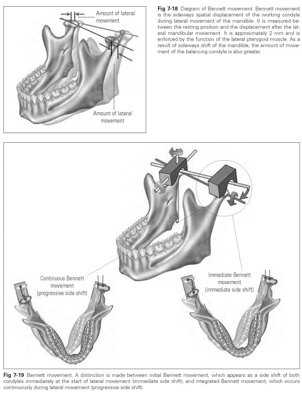
Immediate side shift (initial Bennett movement) means that both condyles are displaced to the side at the start of lateral movement of the mandible; ie, the whole mandible performs a sideways movement running parallel to the hinge axis before the nonworking condyle moves forward, downward, and inward.
This side shift can only be performed with difficulty as an isolated movement, and it may then be interpreted as evidence of joint damage, eg, capsule or ligament strain.
Bennett movement takes place in the simplest case as a linear side shift of the working condyle. In many cases, the condyle is displaced in a particular spatial direction. The spatial side shift is described as follows:
- Laterotrusion is the linear sideways shift of the working condyle to the laterotrusive side.
- Lateroretrusion is the spatial shift of the working condyle to the side and backward.
- Lateroprotrusion is the spatial shift of the working condyle to the side and forward.
- Laterodetrusion is the spatial shift of the working condyle to the side and downward.
- Laterosurtrusion is the spatial shift of the working condyle to the side and upward.
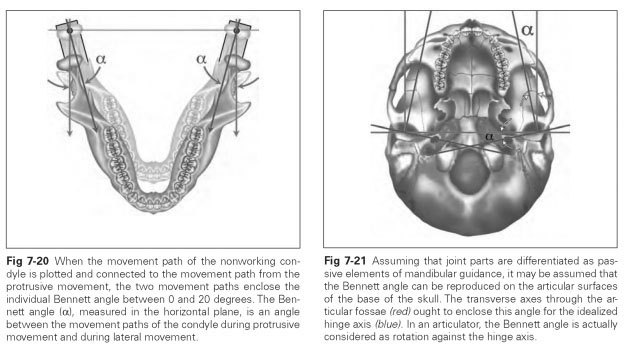
The size of the Bennett movement can be measured directly or expressed indirectly via movement tracings based on the Bennett angle.
The Bennett angle is an angle between the movement paths of the condyle during protrusive and lateral movement of the mandible, measured in the horizontal plane. In a protrusive movement, both condyles slide downward and forward on the condylar paths. When this movement path is plotted, two lines emerge that are roughly parallel to each other (parallels to the median plane) (see Fig 7-12). When the movement path of the nonworking condyle is plotted and connected to the movement path from the protrusive movement, the two movement paths enclose the individual's Bennett angle between 0 and 20 degrees (Fig 7-20). However, the Bennett angle is not the same on each side in humans.
What is the connection between Bennett movement and the Bennett angle? If, during a lateral movement of the mandible, the working condyle were only to complete a rotation around its vertical axis, a Bennett angle of approximately 6 degrees could be measured. Lateral displacement of the Bennett movement will result in an average Bennett angle of about 15 degrees.
Study of the TMJ revealed that the transverse axes of the condyles are not aligned but are rather at an angle to each other and meet at a point in front of the occipital foramen; the same is true of the transverse axes of the articular fossae. Because the condylar paths have differentiated according to the forms of mandibular movement based on the relationship between form and function, it ought to be possible to reproduce the Bennett angle on the articular surfaces of the base of the skull (Fig 7-21). If, for an idealized hinge axis, the transverse axes are drawn through the articular fossae and meet in front of the occipital foramen, this produces an angle between the idealized hinge axis and the transverse axes that roughly corresponds to the Bennett angle. Within the same angle, it can be seen that the hinge axes of the two condyles differ from an idealized, common hinge axis.