Dental Tissues
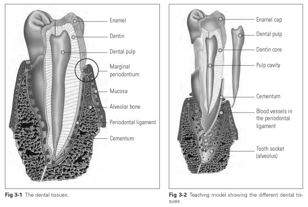
The individual tooth is composed of five different tissue layers, which can be seen without magnification in a cross section of a tooth (Figs 3-1 and 3-2). These tissues can be divided into soft and hard substances:
Hard substances
- Enamel (enamelum)
- Dentin (dentinum)
- Cementum
Soft substances
- Dental pulp (pulpa dentis)
- Periodontal ligament (desmodontium)
The whole tooth with its root is fixed in a bony socket in the jaw, the alveolus. Because the
periodontal ligament is connected to both the cementum of the root and the alveolar bone, a cross section reveals the junction between the
alveolar bone and the periodontal ligament; the junction between the
tooth and the gingival tissue is also evident.
Development of Dental Tissues
The development of dental tissue starts in the fifth week of embryonic life and is not complete until the twentieth year of life, with the formation of the third molars. The developmental processes are genetically controlled and are the same for all teeth. The development of dental tissue is known as odontogeny and starts when the embryo is roughly 9 mm in size. The side swellings for the nose and the primary palate are already identifiable, while there is still a shared nasal and oral cavity. The germ cells for the primary teeth are present in the swellings in the jaw.
The tooth germ cells arise from proliferation of the epithelial cells in the primary jaws and cells from the neural crest in the jaw swellings during weeks 8 to 17 of gestation. During this period, the proliferation of epithelial cells first gives rise to a dental bud and then a cap, and finally, as a result of the high rate of cell division, the tooth caps enlarge into a bell shape, which has roughly the same form as the eventual tooth. In this stage of development, the cells differentiate into the enamel, dental papilla, and follicle (Fig 3-3).
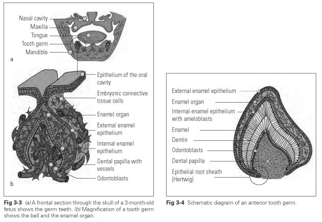
The germ cells of the primary teeth reach the bell stage in week 17 of embryonic life. The permanent first molars reach this developmental stage by week 24, and the permanent second molars reach the bell stage at 6 months after birth. The third molars do not reach the bell stage until 6 years after birth. The bell stage also sees
the start of dentin and enamel formation for the primary and accessional germ teeth. The succes-sional germ teeth develop from the fifth month of pregnancy through to the third year of life.
The successional germ teeth are located lingual to the primary teeth in the bell stage, and then they move to below the roots of the primary teeth when these erupt. This means the anterior germs lie exactly below the tips of the roots, and the premolars are between the splayed roots of the primary molars.
The enamel organ forms the bell with four functionally distinct layers: the external and internal enamel epithelium, the stellate reticulum, and the stratum intermedium.
The enamel epithelium covers the whole surface of the enamel organ (Fig 3-4). The external enamel epithelium forms the outer limit of the enamel organ and, with the cervical loop, is continuous with the internal enamel epithelium at the edge of the bell. The cells of the stellate reticulum make up the inner part of the organ; the cells of the stratum intermedium overlie the internal epithelium as a thin layer. The internal enamel epithelium is the inner layer of the bell and marks the separation from the cells of the dental papilla.
The dental papilla is an accumulation of cells arising from embryonic connective tissue and migrated nerve fibers, which is enclosed by the epithelial bell. A thin layer of connective tissue known as the follicle surrounds the dental papilla and enamel organ.
The cells of the dental papilla control formation of the root and the tooth shape. The enamel-forming cells differentiate from the cells of the internal enamel epithelium, while the dentin-forming cells are differentiated from the peripheral cells of the dental papilla.
The odontoblasts secrete the dentin matrix toward the interface with the internal enamel epithelium and thereby stimulate the enamel-forming cells to produce enamel. The formation
of dentin transforms the organic shape of the bell into a stable molded shape, against which the dental enamel is deposited.