Dentition in the Sagittal Plane
In the sagittal plane, the rows of teeth can be described in their state of intercuspation. Different views of the teeth become visible, depending on which sagittal plane is chosen: The medial plane shows the rows of teeth from the lingual; if the sagittal plane lies outside the dental arches, the rows of teeth are seen from the vestibular view. The intercuspation of the teeth can be seen in each view and follows a regular pattern.
The antagonists rule (occlusion of cusps, fossae, and marginal ridges) denotes the regular in-tercuspation of the rows of teeth, whereby each tooth has two antagonists (Fig 5-20). The mandibular central incisors and the maxillary third molars (or second molars in the absence of third molars) are the exception to the rule, with only one antagonist each.
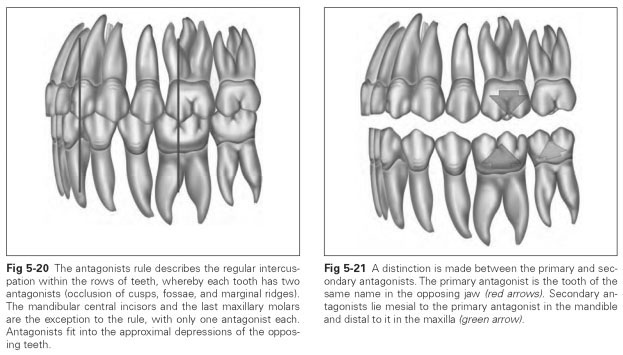
The antagonist (opposing tooth) denotes the tooth that occludes with the corresponding teeth in the opposing jaw when biting down. A distinction is made between the primary and secondary antagonists. The primary antagonist is the tooth of the same name in the opposing jaw. Secondary antagonists lie mesial to the primary antagonist in the mandible and distal to it in the maxilla (Fig 5-21).
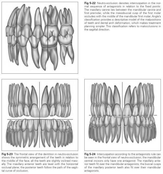
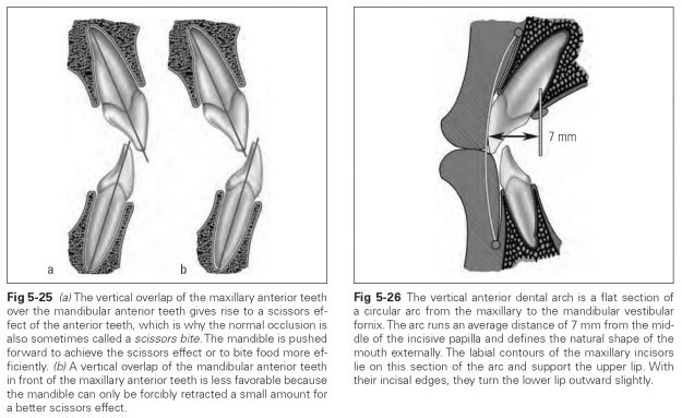
Neutro-occlusion denotes the state of intercus-pation in the normal occlusion, when the tip of the maxillary canine lies between the mandibular canine and the first premolar, while the mesio-buccal cusp of the maxillary first molar fits into the buccal fissure of the mandibular first molar (Figs 5-22 to 5-24). In the medial plane, neutro occlusion displays the regular intercuspation of the anterior teeth in the form of the normal (or scissors) occlusion. The horizontal overlap and protrusion of the maxillary teeth in relation to the mandibular anterior teeth can be seen here; this is also known as the overjet. The scissor effect of the incisors is achieved by the mandible being pushed forward when biting food (Fig 5-25).
The vertical anterior dental arch describes the vestibular angulation of the anterior teeth. Viewing tooth position and inclinations of the alveolar ridge in the medial plane, a circular arc can be drawn from the maxillary to the mandibular vestibular fornix, the contour of which runs 7 mm from the middle of the incisive papilla. The labial contour of the maxillary anterior teeth lies on this arc, so that these teeth support the upper lip with their labial surfaces and the bottom lip with their incisal edges (Fig 5-26). For prosthetic work, the position of the anterior teeth should be reconstructed in keeping with the vertical anterior dental arch.
The vestibular inclination of the anterior teeth can be interpreted as a rudimentary snout formation of the dentition. The protruded part of the mouth allows the teeth to bite food more efficiently without requiring so much force because of the advantageous effect of the scissor action of the relatively sharp incisors. The canines and premolars are used for biting very tough food.
The sagittal plane reveals yet another characteristic form of the dentition: The mandibular teeth are inclined mesially on the jaw, as are the maxillary anterior teeth and premolars; only the maxillary first molar stands almost vertically, while the subsequent maxillary molars appear to be tipped distally (Fig 5-27). This means that the teeth form a curve in the line in which they occlude (Fig 5-28).
The sagittal curve of occlusion (occlusal line) is the continuous connecting line along the occlusal surfaces of the posterior teeth in the maxilla and the mandible in the sagittal direction. It starts at the tip of the mandible, falls away caudally, reaches its lowest point at the first molar, and rises again with the last molars; the distobuccal cusp of the second molar lies at the same level as the tip of the canine. If the sagittal curve of occlusion is drawn along both the mandibular and maxillary buccal cusp tips and is projected onto the sagittal plane, the two lines run parallel, but the maxillary curve is displaced caudally in comparison with the mandibular curve, according to the depth of intercuspation or cusp height. The sagittal curve of occlusion divides the occlusal plane at four common points: on the right and left at the distobuccal cusp of the second molar and at the tips of the right and left canines. If the path of the occlusal plane is known, the sagittal curve of occlusion can be reconstructed.

The curve of Spee (named after Count Ferdinand Spee) is a sagittal curve of occlusion that bends sharply downward, its dorsal projection touching the anterior surface of the condyle (Figs 5-29 and 5-30). The tooth axes are inclined so that their projections meet in the orbital cavity as radii of the curve of Spee. According to Spee, the forward movement (protrusive excursion) of the mandible runs along this curve, with all the teeth maintaining sliding contact. This is not true of the normal occlusion. The sagittal curve of occlusion follows a flatter course and enables separation of the posterior teeth during protrusive excursions.