Mucous Membrane
The mucous membrane (mucosa, tunica mucosae) forms the epithelial lining of the inner surface of the digestive tract (intestinal mucosa), oral and nasal cavity, airways, respiratory organs, and urinary and sexual organs.
Layers of the mucous membrane
The mucosal epithelium (epithelium mucosa) is mostly nonhorny squamous epithelium or ciliated epithelium, which is constantly kept moist and slippery by a mucous secretion made up of mucus cells (goblet cells) or by mucous glands. The mucus also acts as a chemical buffer. The oral cavity is lined with multilayered squamous epithelium, the intestine with single-layered prismatic epithelium, the respiratory tract with multiple rows of ciliated epithelium, and the urinary tract with transitional epithelium.
Mucosal connective tissue (lamina propria) lies directly below the mucosal epithelium and ensures that substances are transported between the surface and the vessels. Blood and lymphatic vessels as well as autonomic nerve fibers run through this layer.
The mucosal muscle layer (lamina muscula-ris mucosae) is only found in the digestive tract, starting in the esophagus and extending to the rectum.
Submucous connective tissue (tela submucosa) is the innermost layer, in which blood and lymph vessels and autonomic nerve fibers are brought closer to the mucous membrane.The other layers are supported on the submucous connective tissue in a free-moving way.
Functions of the mucous membrane
The functions of the mucous membrane include the following:
- Protective function: In this case, effective protection against bacteria, viruses, and parasites; however, the mucous membrane also ensures mechanical and chemical protection as well as biologic protection. These functions are mainly performed by the multilayered squamous epithelium and ciliated epithelium.
- Sensory function: In this case, perceiving pain in response to temperature differences and mechanical effects.
- Production of mucus: As a transporting mucus or as glandular products containing digestive enzymes.
- Absorptive function: Absorbing nutrients in the digestive tract. These functions are undertaken by highly prismatic epithelial cells.
Mucous membranes do not have any hairs or sweat glands, while sebaceous glands are only found in the transitional epithelium of the lip mucosa. Generally the mucosa is free-moving on its connective tissue support. The exception is the oral mucosa, which is fixed in a (relatively) immobile way onto the bony jaws, the mucosal epithelium being interwoven with the mucosal connective tissue. Here the connective tissue lamina propria functions as an adhesive layer for the mucosal epithelium.The mucosa is normally non-horny, but a horny layer (callosity) may develop at mechanically stressed sites.
The mucosa of the oral cavity is made up of multilayered, nonhorny squamous epithelium, which is translucent. Except in the gingiva (the mucosal area of the alveolar ridges as far as the teeth) and the area of the anterior hard palate, a large amount of mixed mucus and salivary glands are located in the oral mucosa. Salivary glands lie in the oral cavity and produce saliva. They can be classified according to their size.
Small salivary glands in the mouth are mucous and serous glands and include the following:
- Labial glands (glandulae labialis)
- Buccal glands (glandulae buccalis)
- Molar glands (glandulae molares), which open into the vestibular area of the mouth
- Palatine glands (glandulae palatinae)
- Lingual glands (glandulae linguales), which open into the oral cavity
Large salivary glands include the following:
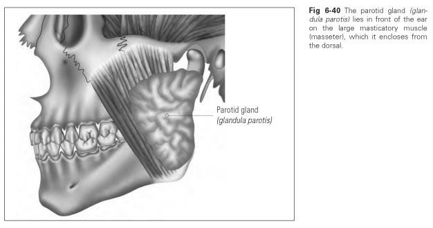
- Parotid gland (glandula parotis), a serous gland that lies in front of the outer ear and exits in the cheek, level with the second molar (Fig 6-40)
- Sublingual gland (glandula sublingualis), a mixed gland that lies in the sublingual fossa of the mandible and exits in the sublingual caruncle, a papilla of mucous membrane next to the frenulum of the tongue (Fig 6-41)
- Submandibular gland (glandula submandibu-laris), a mixed gland that lies in the mandibular fossa of the mandible and also exits in the sublingual caruncle (see Fig 6-41).
All of these glands perform an important metabolic function and produce a specific secretion that altogether make up saliva.
