Muscles of Mandibular Movement
The TMJs and teeth are the rigid guide elements of mandibular movement; muscle guidance forms the third element. Muscles of mastication, as an active part of the locomotor system, move the mandible against the cranium and produce masticatory pressure. The TMJ is relatively elastic and unstable because of the intrinsic mobility and compressibility of tissue and the bisection of the joint by the disc. Tooth guidance functions within small deflections of movement (ie, only movements with tooth contact are maintained). All movements outside of tooth contact are mainly guided by the interaction of the different masticatory muscles. Each person develops a differentiated habit of masticatory movements for different food consistencies.
The habitual movements are matched to the shapes of the teeth and the form of the TMJ. Both tissue parts "imprint" themselves onto this movement habit, just as this habit is determined by the particular form of the other guidance factors. The process of differentiation follows the laws of form and function and is always changeable; ie, movement habits and the TMJ adapt to changes within the dentition, whether due to tooth loss or prosthetic treatment. However, this means that faulty dentures can lead to pathologic changes in these guide elements.
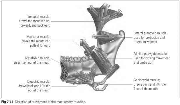
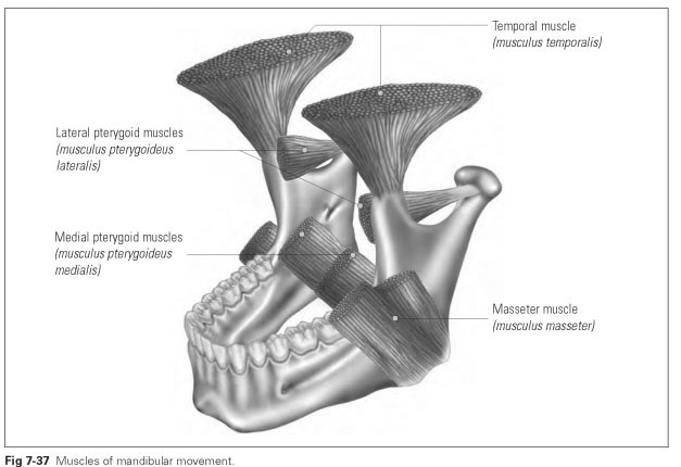
The masticatory musculature does not include all the muscles involved in mandibular movement but only those that are excited by the mandibular branch of the trigeminal nerve and extend from the skull to the mandible. They form a functional unit that developed genetically from the branchial muscles (branchial = relating to fishes' gills). There are four pairs of muscles that originated from a common muscle mass and are still interconnected at their borders. The four paired masticatory muscles are the temporal muscle, the masseter muscle, and the medial and lateral pterygoid muscles (Fig 7-37).
A theoretical classification of muscles in relation to mandibular movement divides them into mouth-closing and mouth-opening muscles and those that pull forward and sideways. This classification names the main functions of these muscles and conceals the relationship whereby mandibular movement is only performed by the combined activity of the different groups of muscles. For instance, all the muscles of mandibular movement become active during highly discriminating tactile movements.
Mouth-closing muscles (masseter, medial pterygoid, temporal muscle) produce an average of 700 N and a maximum of 1,250 N of masticatory pressure in the molar region. In the incisal area, masticatory pressure is approximately 400 N because of the unfavorable leverage conditions. Where a denture is not supported by the periodontium, the average masticatory pressure is about 100 N.
Mouth closing (elevation; raising the mandible) is performed by the masseter muscles, medial pterygoid muscles, and the vertical fibers of the temporal muscles; the suprahyoid muscles prevent closing from occurring too quickly or from being uncontrolled.
Mouth-opening muscles are muscles of the floor of the mouth, namely the mylohyoid, geniohyoid, and digastric muscles as well as the suprahyoid and infrahyoid muscles. Sideways and forward movement is performed by several muscles.
Mouth opening (depression; lowering the mandible) is performed by the geniohyoid and mylohyoid muscles, the anterior bellies of the digastric muscles, and parts of the lateral pterygoid muscles. In this movement, the hyoid is fixed by the infrahyoid muscles.
Forward movements of the mandible (protrusion) are effected by the lateral pterygoid muscles, the transverse fibers of the medial pterygoid muscles and masseter muscles, as well as the fibers of the temporal muscles that run forward.
Sideways movements (laterotrusion; grinding movements) are firstly performed by the muscles on the opposite side, with support from the backward-running fibers of the temporal muscle on the laterotrusive side. When the mandible is sliding into centric occlusion from the lateral position, all the muscles on the laterotrusive side come into action, so that the mandible is also pulled forward from a slightly retruded position.
The mandible is pulled backward (retrusion) by the horizontal fibers of the temporal muscles and the infrahyoid muscles, the hyoid bone being fixed by the infrahyoid muscles.
Figure 7-38 illustrates the direction of movement of the masticatory muscles.