Orthodontic Model Analysis
Local occlusal deviations, malpositions of teeth, and anomalies in occlusal position can be identified directly in the patient or through the use of orthodontic models. These models are excellent aids to a thorough analysis of an occlusal anomaly and for planning purposes. As a means of measuring the shape of the dental arches and describing the deformations in the arches, these models are even better suited than examination of the patient.
There are several methods of model analysis with which a relationship between tooth width and dental arch dimensions can be mathematically recorded. A specific width and height of dental arch pertains to a particular width of incisors (Fig 10-3). In statistical records of normal dental arches, this ratio is summarized in formulae and tables.These analysis methods provide a good indication of the dental arch widening or narrowing required.
Pont's index is the best-known method of model analysis, in which the sum of the width of all four maxillary incisors (sum of incisors = SI) indicates the ideal width of the dental arch at two points, namely at the first premolars and the first molars.
To determine the anterior arch width, Pont's index uses the following equation:
(Sl x 100)/80=P-P
where P is the first premolar.
To measure the posterior arch width, Pont's index uses the following equation:
(Sl x 100)/64 = M-M
where M is the first molar.
For the length of the dental arch, the vertical distance from the anterior arch width to the incisors is calculated as follows:
Maxillary dental arch length = (Sl*100)/160
Mandibular dental arch length = (Sl*100)/160 - 2
It becomes clear that the position of the measuring points on the teeth must be precisely located for the measurement to be meaningful and especially verifiable. The sum of incisor widths is formed from the individual measurements of the mesiodistal distances between the approximal contacts in the four maxillary incisors. If the maxillary tooth widths cannot be measured because they have not all erupted, the widths of the mandibular incisors should be measured. Because there is a fixed size ratio between the mandibular and maxillary incisors, Pont's index can be calculated by means of this ratio. On average, the ratio of the maxillary incisor widths to the mandibular incisor widths is 4:3.
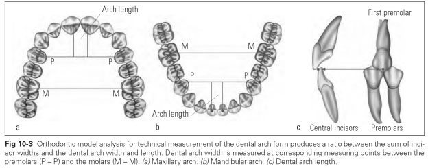
The measuring points for the anterior arch width on the maxillary first premolars lie in the middle of the central developmental groove. The measuring points for the posterior arch width on the maxillary first molars lie in the central fossa. In the mandible, the measuring points of the anterior arch width lie at the contact point between the first premolar and second premolar. The posterior arch width in the mandible runs between the central buccal cusps of the first molars. Orthodontic dividers can be used to measure the arch widths intraorally (Fig 10-4).
The maxillary and mandibular dental arch lengths run perpendicular to the anterior arch width to the occlusal contact points of the central incisors; this means they lie on the incisal edges of the mandibular central incisors and on the palatal surfaces about 2 mm from the incisal edges of the maxillary central incisors.
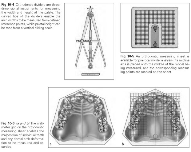
For measuring orthodontic models, a transparent sheet bearing a grid marked out in centimeters and millimeters is available. Pins are often placed at the midline, with which the measuring sheet can be fixed to the midline of the model (Fig 10-5). Measurement of the maxillary dental arch involves the following steps:
- Measure the incisor widths.
- Calculate the anterior and posterior arch widths as well as the anterior arch length.
- Establish and mark the middle of the model.
- Place the measuring sheet with the midline mark over the middle of the model.
- Lay the measuring sheet through the assumed anterior arch width.
- Read off deviations from the actual values in two directions and note.
Asymmetries of the dental arches and migration of individual teeth are thus identified (Fig 10-6). The system error with this model analysis lies in the natural variance of individual dentitions and in the uncertainty of the position of the fixed points from which the deviations are determined. If the reference points (distal premolar or raphe-papillary transversal) are displaced, only relative deformations or displacements can be ascertained. Extraoral fixed points such as cranial reference lines, which are fixed in their direction and their distance from intraoral fixed points, allow more objective measurements to be made.