Types of Joints
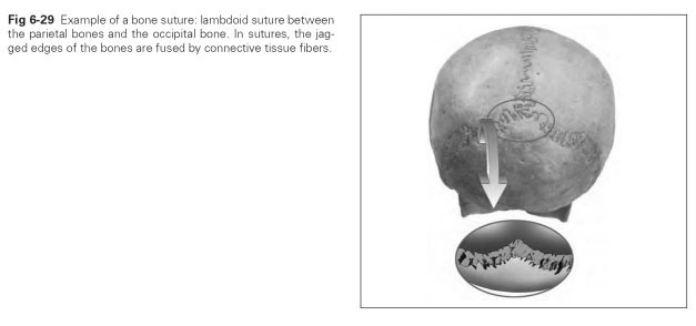
A distinction is made between joints (junctions or juncturae) and true joints (synovial joints or articulations) in terms of the way bones are joined together. Juncturae are the solid bony connections such as sutures (Fig 6-29), synchondroses (cartilaginous joints), and ossified connections. These are defined according to the nature of the connecting material:
- The fibrous joint (junctura fibrosa) denotes two bones united by connective tissue fibers in the bone sutures; the two bones are "sewn together" by fibers (eg, the cranial sutures).
- The cartilaginous joint (junctura cartilaginea) denotes two bones united by fibrocartilage, such as the symphysis of the pelvic bones.
- The osseous joint (junctura ossea) can develop from the ossification of a fibrous joint. Examples of this type are cranial sutures, the symphysis in the mandible, and the sacral vertebrae.
- True joints or articulations (juncturae synovia-lis, articulationes) permit the movement of two bones against each other; they can be divided into freely movable (diarthroses) joints and rigid joints (amphiarthroses). The rigid joints with a fixed joint capsule and short, strong ligaments permit only slight mobility or simply have a cushioning effect (eg, the tarsals and carpals of the feet and hands).
Structure
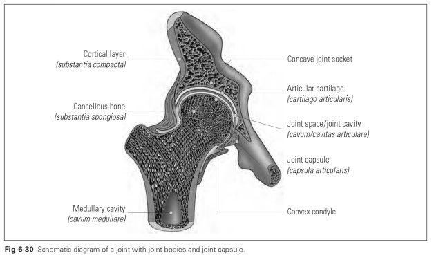
Joint bodies at the ends of the bones that are to be connected bear the articular surfaces with a 3-to 4-mm-thick layer of hyaline or fibrocartilage as the articular cartilage (Fig 6-30). Articular surfaces can be divided into the convex condyle and the concave socket. The purpose of the articular cartilage is to achieve considerable closeness of fit, to act as a shock absorber due to its elasticity, and to save the bone from splintering in response to mechanical strain. It is noticeable in some joints that the surfaces do not fit together exactly; in other words, they display incongruence that is compensated for by articular discs (eg, meniscus articularis in the knee or discus articularis in the TMJ).
The joint capsule (capsula articularis) is made up of a strong coating of connective tissue that provides an airtight seal to the joint cavity. The joint capsule is fixed to both articulating bones and is made up of two layers. The outer fibrous layer (membrana fibrosa; fibrous membrane) consists of crosswise collagen fibers that are reinforced by ligaments. The inner layer (membra-na synovialis; synovial membrane) is a thin layer through which blood vessels and nerves pass and whose processes, known as synovial folds or villi, secrete the synovial fluid.
Synovial fluid (synovia) is a tough, stringy substance made up of tissue fluid, dead epithelial cells, and other cell fragments that reduces friction between the joint surfaces like a lubricant.
A joint cavity (joint space; cavum articulare) is a capillary space in a living joint that is squeezed together by external tissue pressure. This joint space is the characteristic feature of a true (synovial) joint.
The synovial bursae protect the tendons against particular stress when they slide over the bones as a result of the movement of the joints. The usual location of these bursae is the tissues of the joint capsule, ie, where tendons attach to the bone in a wide angle and where strong shearing movements against the skin and bone take place.
Tendon or synovial sheaths are bundles of collagen fibers that extend between the tendons running in different directions to ensure that the tendons are guided alongside each other. Connective tissue membranes are also found enclosing long tendons, guiding the tendons and carrying vessels that feed the tendons.
The mobility of the joints depends on the shape and closeness of fit of the articular surfaces (bone guidance), the arrangement and strength of the articular ligaments (ligament guidance), and the arrangement and functional direction of the muscles acting on the movable bones.
The particular geometric shape of the articular surfaces determines the direction of motion of a joint, ie, the number of degrees of freedom.There are six basic types—hinge, ellipsoidal, spheroidal
(or ball-and-socket), saddle (or sellar), pivot, and plane—depending on the geometric shape and the principal directions of motion (Fig 6-31).
The hinge joint (or ginglymus) permits rotation around one axis, which lies perpendicular to the movable parts. A hinge joint has only one degree of freedom. The knee and the interphalangeal joints in the hands are examples of hinge joints.
The ellipsoidal joint permits movement around two axes of rotation (biaxial). The condyle is eggshaped (ellipsoid) and fits into the matching joint socket. Examples of this type are the joints between the wrist and the forearm bones and the joints between the occiput and the first vertebrae.
The spheroidal or ball-and-socket joint has three degrees of freedom; ie, movements are possible in all spatial axes. Examples are the hip and shoulder joints.
The saddle or sellar joint permits movements around two spatial axes, which means it has two degrees of freedom as a result of two arched articular surfaces with matching curves. One of the joints in the thumb is an example of this type.
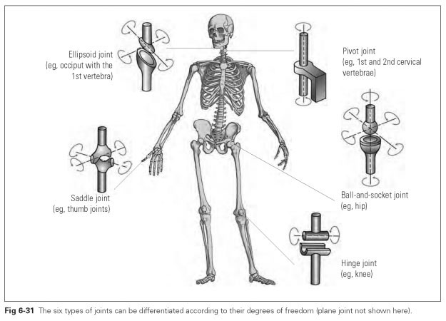
The pivot joint occurs when a ring-shaped bone surrounds a peg or pivot of bone, making rotation around the axis of the pivot possible. Both the bony ring and the peg of bone are able to rotate in this case. The joint between the first and second cervical vertebrae (atlanto-axial joint) is an example of a pivot joint.
The plane joint permits a sliding movement by the flat articular surfaces against each other.