Bones of the Cranial Vault
The frontal bone (os frontale) arises from a paired arrangement where two parts are fused into one bone in the adult (Fig 6-9).The frontal bone forms the anterior area of the cranium and the roof of the orbits. Behind the prominent orbital swellings are the frontal sinuses, which are air-filled and lined with mucous membrane. These sinuses are connected to the actual nasal cavity as paranasal sinuses. The nasal part (pars nasalis) of the frontal bone, on which the nasal skeleton grows, lies between the two roofs of the orbits.
The occipital bone (os occipitale) forms the back of the head (the occiput) and the bony casing for the cerebellum and parts of the cerebral hemispheres (Fig 6-10). The occiput is where the muscles of the neck attach. The large occipital foramen (foramen occipitale) at the base of the skull is the opening through which the spinal cord, located in the spinal canal, passes. The articular eminences at the front next to the occipital foramen are the joint surfaces of the atlanto-occipital and atlanto-axial joints. The occipital bone comprises four elements that are grouped around the occipital foramen and form one unified bone: at the front, the unpaired piece of bone called the basilar part (pars basilaris); at the sides, the lateral parts (partes laterales); and, at the back, the occipital squama (squama occipitalis).
The parietal bone (os parietale) forms the roof of the skull in its middle section and is hence the superiormost aspect of the cranium. The sutures to the adjacent bones, the frontal bone, the occipital bones, and between the parietal bones themselves ossify very late and thus enable the skull to enlarge.
The sphenoid bone (os sphenoidale) is a symmetric bone at the base of the skull (Fig 6-11), from whose transverse bodies run two horizontal pairs of wings (alae majores and alae minores) and a vertical pair of wings, the pterygoid processes (processus pterygoidei; Greek, pteryx = wing).
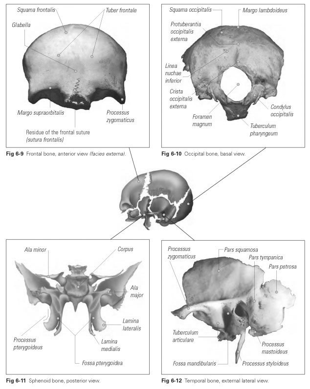
Pterygoid processes can be divided into one lateral and one middle lamella, which serve as the points of origin for the pterygoid muscles for mandibular movement.These pterygoid processes are joined to the infratemporal surfaces of the body of the maxilla and contact the vertical plates of the palatine bones. The greater palatine canal
(canalis palatinus major) is formed by these three bony parts and opens into the palatine foramina. Blood vessels and nerve pathways run through the canal into the posterior palatine region.
The two horizontal pairs of wings contain important openings through which nerves from the oral cavity and the orbits pass. The optic canal (canalis opticus) is located in the small wing (ala minor) and allows the optic nerve to pass through from the cranial cavity to the orbit. The large wing (ala major) is the site of the round foramen (foramen ro-tundum), a roundish canal that carries the second branch of the trigeminal nerve (maxillary branch). The oval foramen (foramen ovale), the oval hole for the third branch of the trigeminal nerve (mandibular branch), is also located in the ala major.
The temporal bone (os temporale) is the part of the skull on which the head mainly rests during sleep (Fig 6-12). It takes its name (literally "time bone") from the fact that the hairs at the temples are the first to go gray, hence indicating the passage of time in life. The structure of the temporal bone is very complex. The temporal bone houses a bony capsule for the organ of hearing and balance (the ear), which is why this bone has numerous cavities and canals. The temporal bone is divided into three parts: the petrous, tympanic, and squamous parts.
The petrous part (pars petrosa) is the posterior region of the temporal bone and is made up of very hard bone. The mastoid process is a bony bulge projecting downward from the petrous part that arises because of the pulling effect of turning the head.
The tympanic part (pars tympanica) forms the external auditory canal (or external auditory meatus) as a tube, at the inner end of which the eardrum is stretched.
The squamous part (pars squamosa) is the actual body of the temporal bone and forms the lateral wall of the skull. The zygomatic process extends forward from this squamous part and, together with the temporal process of the zygomatic bone, forms the zygomatic arch (arcus zygo-maticus). On the underside of the temporal squama lies the socket (fossa mandibularis) for the TMJ, which is bounded anteriorly below the root of the zygomatic process by the articular tubercle (tuberculum articulare). The styloid process (processus styloideus), which can vary in length, is the bony attachment for the stylomandibular ligament.