Base of the Skull
The separation of the skull into the facial part and the cranial vault can be seen from the outer surface of the base of the skull (Fig 6-3). Removing the mandible to view the base of the skull (basis cranii) reveals the very highly differentiated anterior facial part and the clearly defined neurocra-nial part. The anterior area is formed by the two maxillary bones, to which the palatine bones are attached at the rear. To either side of that are the zygomatic bones (zygomata), which join the temporal bones to form the zygomatic arch (arcus zy-gomaticus). The middle connection between the roof of the palate (palatine vault) and the body of the sphenoid is formed by the vomer; the lateral connections between the sphenoid and the maxilla are formed by the pterygoid processes of the sphenoid bone.
The area of the cranial vault, formed by the sphenoid bone, the temporal bones, and the occipital bone, contains several holes and fissures as openings through which blood vessels and nerves pass.The large occipital foramen (foramen occipitale magnum), where the articular eminences of the occipital condyles (condyli occipitales) are located anterolaterally, is noticeable. The joint surfaces of the TMJs are found on the temporal bones, right at the roots of the zygomatic arch near the external auditory orifices.
The inner surface of the base of the skull (Fig 6-4) is smooth with delicate depressions formed by the cerebral gyri (impressiones digitatae). Fine sulci and holes can also be seen as well as canals that can be traced as the course of blood vessels and nerves.
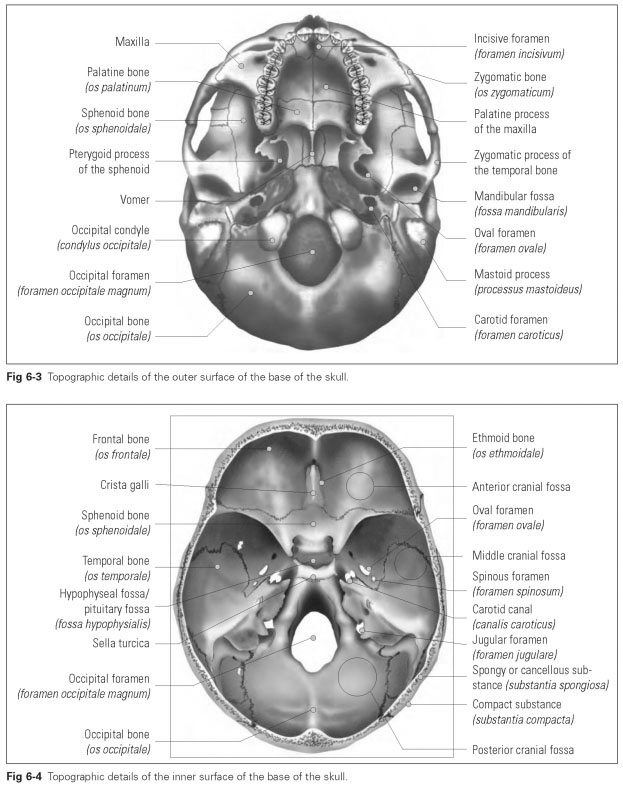
Three fossae can be seen on the inside of the base of the skull: an anterior fossa, middle fossa, and posterior fossa. The anterior cranial fossa (fossa cranii anterior) is mainly formed by the frontal bone, which lets through a sharp lamella of the ethmoid bone in the middle. The floor of the anterior cranial fossa is made up of extremely thin bony plates that form the roof of the orbits and, anteriorly and vertically, the walls to the frontal sinuses.
The middle cranial fossa (fossa cranii media) is halved in the middle of the skull by the sella turcica of the sphenoid bone. The sphenoid and temporal bones form the base of the middle cra-
nial fossa.This middle area of the base of the skull is where the most openings for nerves and blood vessels are found.
The posterior cranial fossa (fossa cranii posterior) contains the striking occipital foramen in the occipital bone, which, with parts of the temporal bones, forms the bony base of the posterior cranial fossa.
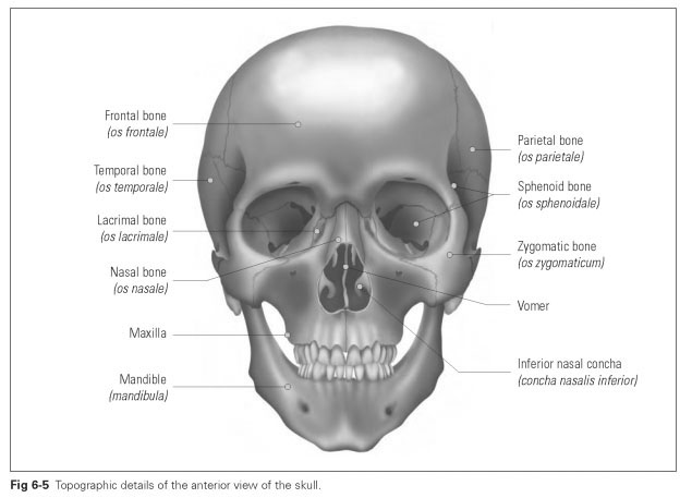
Figures 6-5 and 6-6 show the topographic details of the skull from anterior and lateral views.
The cranial sutures join together the bones of the cranium. In the cranial sutures, the bones are indented and contain narrow strips of connective tissue, which give the bony vault of the skull a certain elasticity. As growth areas, the sutures allow cranial growth up to the age of 40 or 50 years.This is because the brain continues growing until this stage of life, which forces the cranium to grow with it. Age-induced ossification of the cranial sutures only happens once the brain has stopped growing.The individual sutures are the following:
- The coronal suture (sutura coronalis) runs crosswise over the skull cap and joins the frontal bone to the parietal bones.
- The sagittal suture (sutura sagittalis) is the suture between the parietal bones and runs from the coronal suture to the lambdoid suture.
- The lambdoid suture (sutura lambdoidea) runs between the occipital bone and the parietal bones; the continuation between temporal bone and occipital bone is known as the occipitomastoid suture (sutura occipitomastoidea).
- The sphenofrontal suture (sutura sphenofron-talis) joins the frontal bone to the wings of the sphenoid.
- The squamous suture (sutura squamosa) joins the parietal and temporal bones to the skull at the sides.
The suture between the two squamae of the frontal bone (sutura frontalis) closes by the end of the sixth year of life; sometimes a fine residual suture is left above the root of the nose. In the skull of the newborn, the cranial sutures, such as the coronal, sagittal, and lambdoid, are permeated by sections of connective tissue and are elastic. The sutures are open in the form of fontanels so that the brain can be seen pulsating beneath (fonticulus; Latin, fons = fountain) (Fig 6-7).
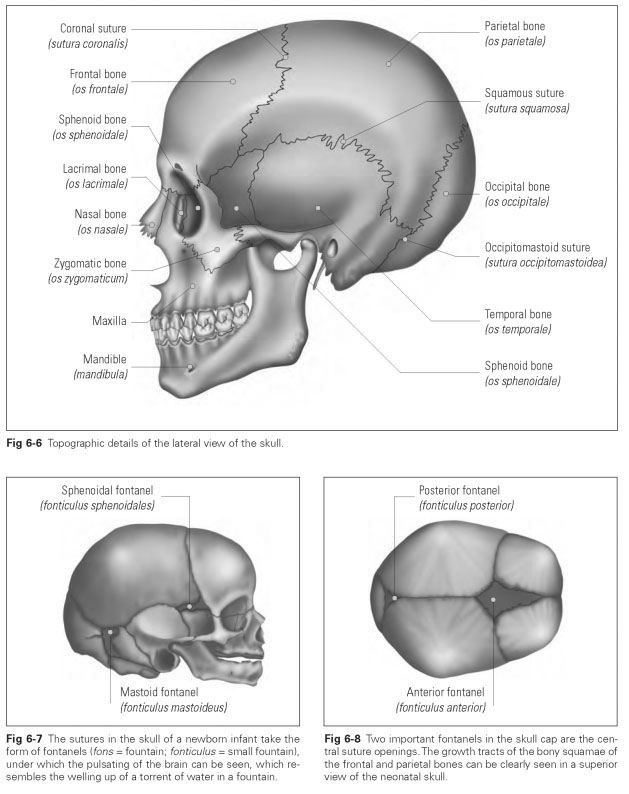
There are two central and two lateral fontanels, with the lateral ones being less important (Fig 6-8). The large, anterior fontanel is formed by the frontal bone and the parietal bones; the posterior fontanel lies at the crossover point with the lamb-doid suture. The fontanels only close fully during the second year of life. In the fontanels, the individual cranial bones can move against each other so that the relatively large skull is able to change shape safely as it passes through the narrow birth canal.