Canines
Dens caninus means dog's tooth (canis = dog); dens angularis from angulus = angle or corner; dens cuspidatus = single-cusp tooth from cuspis = cusp, protuberance.
The canine resembles a fang in dogs, which is the origin of its name. The maxillary canine is often popularly known as the "eye tooth" because inflammation affecting the canine root (periapical processes) causes swelling around the eyes.
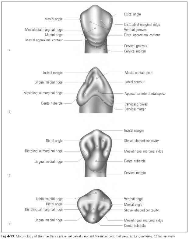
The maxillary canine (dens caninus maxillae) (Figs 4-33 to 4-35) has a long, sturdy root with a stress-resistant periodontium. The root apex is slightly curved distally, reflecting the root characteristic. The pulp cavity widens in the coronal region. Viewed approximally, it is noticeable that the labiopalatal diameter is largest at the cervix. This gives the tooth its statically favorable chisel shape.
The maxillary canine is an excellent abutment and clasp tooth for partial dentures because of its position and its sturdy shape. It should be retained as long as possible. Despite resection of the root apex (apicectomy), ie, the tooth being nonvital, it can still be retained as a load-bearing abutment for a long time (eg, in a partial denture construction). Its periodontium is particularly stress resistant because a large number of fibrous bundles are arranged crosswise and radially, securing the tooth against transverse masticatory forces and torsion. It serves to grasp (and tear) food. The canine is a rudimentary fang that has regressed as a result of functional adaptation; thus, the canine and the premolars are instinctively used to bite tough food.
The average dimensions of the maxillary canine are:
- Incisal crown width (mesiodistal): 7.6 mm
- Cervical crown width (mesiodistal): 6.0 mm
- Crown depth (labiolingual, cervical): 8.0 mm
- Crown length: 11.0 mm
- Total apicocoronal length: 27.0 mm
The vestibular surface (see Fig 4-33a) exhibits the striking angular form: The cutting edge is made up of two sides of differing length inclined toward each other. The mesial side is shorter and does not recede as steeply as the longer distal side. The transitions between the cutting edge and the approximal surfaces thus lie at different heights:The mesial edge is shifted incisally, while the distal edge is displaced in a cervical direction; the mesial contact point is displaced more toward the incisal. One angle characteristic can be identified because the distal transition of the incisal margin is clearly rounded, unlike the sharp-edged mesial transition.
From the tip of the incisal edge, the sturdy medial ridge runs cervically as it changes into the prominent transverse convexity of the cervix. Poorly developed cervical grooves are found here. The medial ridge divides the labial surface into a narrow mesial and a broad distal facet. The horizontal curvature of the canine is strongly developed, with both facets receding from the central ridge to the adjacent teeth. Both facets contain a distinct marginal ridge in the vertical direction.
The neck of the tooth is arched and contains the strong vertical curvature to protect the marginal periodontium. The approximal edges run closely together from the contact points in a cervical direction; in the middle, the distal approximal edge is rather concave centrally, whereas the mesial edge runs virtually straight.
The mesial approximal surface (see Fig 4-33b) exhibits the pronounced wedge shape of the ca-nine.The mesial incisal edge lies inferior to the tip of the tooth. The vertical convexities of the vestibular and lingual surfaces that protect the marginal periodontium can be seen. While the vestibular convexity runs evenly incisocervically, the lingual surface in the cervical third bends inward and only achieves the outer convexity through the tubercle. The heavy tubercle gives the tooth its bulky appearance. The cervical margin curves in an incisal direction. The tip of the canine lies centrally in relation to the base of the crown. Weak cervical grooves can be seen labially.
The lingual surface (see Fig 4-33c) is smaller than the labial surface but with the same basic triangular shape. The tubercle is strongly developed. The marginal ridges are very prominent, as is a central ridge starting from the tubercle. This ridge is described as the canine guidance ridge because it is here that a certain guidance of the opposing teeth (antagonists) takes place during mandibular movement. The distal marginal ridge is developed into a strong masticatory edge incisally. The central ridge develops cusplike into the incisal tip, which approximates to a masticatory surface in the distal portion of the crown. The cervical line is arched, and the tooth bulges out below the line to protect the gingiva.
The incisal view of the maxillary canine (see Fig 4-33d) shows the strongly developed curvature characteristic, ie, the mesial facet is narrower than the distal, and both facets recede laterally, following the curvature of the dental arch. The strong medial ridge can be seen labially, and the cusplike tubercle is visible palatally.
The approximal surfaces are acutely angled mesially and slightly curved distally. A gentle internal curvature of the distal approximal surface serves as the contact area to the premolar. The incisal margin is curved in line with the curvature characteristic and in the distal portion is wider than the masticatory edge.
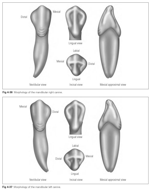
The mandibular canine (dens caninus man-dibulae) (Figs 4-36 and 4-37) resembles the maxillary canine in all characteristics; in terms of form and function, the canines bear the closest resemblance to each other of any teeth. However, the mandibular canine is much more slender and narrower than the maxillary canine, both in the crown and in the root. It has a stronger angle characteristic, with the distal transition from the incisal margin to the approximal surface located more apically than the mesial. Its root is not only much shorter, but in some cases it can be divided: The tooth may even become two-rooted. The stronger horizontal curvature characteristic is also evident; the canine tip generally is in line with the middle of the crown base. The tip of the mandibular canine abrades the tubercle and palatal medial ridge of the maxillary canine, and the teeth show corresponding wear facets.
The average dimensions of the mandibular canine are:
- Crown width (mesiodistal): 6.4 mm
- Crown depth (labiolingual): 7.8 mm
- Crown length: 11.4 mm
- Total apicocoronal length: 25.4 mm
The vestibular surface shows the typical canine shape but is narrower at the contact points in comparison with the maxillary canine; the approximal edges do not run parallel.The mesial incisal margin is shorter and higher than the distal margin, which also recedes more sharply than in the maxillary canine. This means that the distal approximal surface is extremely small. The medial ridge, marginal ridges, vertical grooves, and cervical grooves are prominent. The horizontal transverse convexity is more pronounced on the mandibular than on the maxillary canine.
The lingual surface is not as strongly developed and is less concave than in the maxillary tooth. There is a weak medial ridge, hardly any marginal ridges, and a very flattened dental tubercle; variations in ridge formation are very rare.
From the mesial approximal view, the crown appears to be inclined lingually. However, the tip of the mandibular canine, like that of the corresponding maxillary tooth, is aligned with the midline of the crown base. The appearance of an incline results from the flattened dental tubercle and the vertical curvature of the labial surface.
The incisal view shows the stronger horizontal curvature of the labial surface.The lingual surface appears to taper considerably, and the approximal surfaces are depressed. The incisal margin is more strongly angled than in the maxillary canine; ie, the mesial edge faces the anterior teeth, while the distal edge is far more curved toward the posterior teeth. The strong development of the labiolingual diameter at the crown base is noticeable.
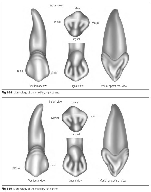
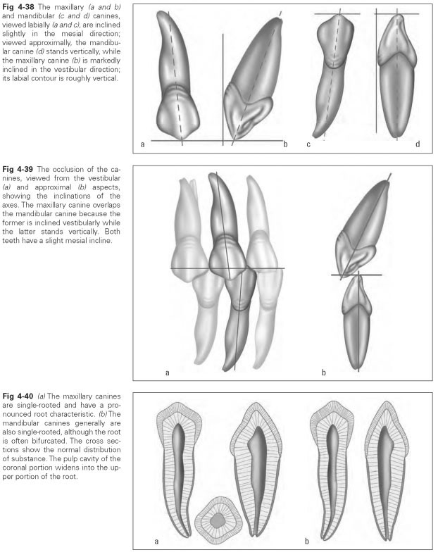
Figures 4-38 to 4-40 show the inclinations of the tooth axes of both maxillary and mandibular canines as well as details of their morphology.