Facial Bones
The ethmoid bone is a sievelike, perforated, air-filled bone with a number of cavities known as ethmoidal cells (cellulae ethmoidales), which feed into the nasal cavity and are part of the paranasal sinuses. A plate of the ethmoid bone projecting downward, known as the perpendicular or vertical plate (lamina perpendicularis), forms the superior part of the nasal septum.The superior edge of the perpendicular plate forms a cockscomblike crest (crista galli) extending into the cranial cavity. A narrow bony plate forms the roof of the ethmoid bone (cribriform plate; lamina cribrosa), which is perforated with numerous holes like a sieve. The lateral walls of the ethmoid bone are smooth, paper-thin bony plates (orbital lamina; lamina orbitalis) that form one part of the medial orbital wall. The ethmoidal labyrinth is located on either side of the nasal septum and, with its curved, twisted lamellae of bone (superior and middle nasal conchae), forms the branched passages of the nasal cavities and the paranasal sinuses.
The superior nasal concha (concha nasalis superior), like the middle nasal concha (concha nasalis media), lies at the medial surface of the ethmoidal labyrinth; from this surface a narrow, hook-shaped uncinate process (processus unci-natus) is projected backward and lies in front of the maxillary hiatus (hiatus maxillaris).
The inferior nasal concha (concha nasalis inferior) is a shell-shaped or turbinate bone that lies along the lateral wall of the nasal cavity and covers part of the opening to the maxillary sinus. At its superior aspect, the inferior nasal concha meets the ethmoidal labyrinth. A process (processus lacrimalis) is directed upward to the lacrimal bone and, together with that bone, forms the medial wall of the nasolacrimal canal.
The nasal bone (os nasale) is a small, rectangular bone that forms the bridge or dorsum of the nose. It attaches to the frontal process of the maxilla and at its superior aspect borders the frontal bone.
The lacrimal bone (os lacrimale) is located at the anterior medial wall of the orbit and contains a vertical depression (the lacrimal sulcus), which with the lacrimal sulcus of the frontal process on the maxilla forms a connecting passage to the nasal cavity.This is where the lacrimal sac is located.
The vomer resembles the blade of a plow and forms the inferior aspect of the nasal septum.The vomer borders on the perpendicular process of the ethmoid bone superiorly and the sphenoid bone posteriorly. The posterior, free edge of the vomer separates the two posterior nares (choa-nae) from each other.
The zygomatic bone or cheekbone (os zygo-maticum) is a rectangular bone that presents two processes: the temporal and the frontal processes. It forms the bony foundation of the cheek. The bone forms a bridge between the facial part of the skull and the lateral walls of the cranium via the zygomatic arch, which is shaped by the temporal process and the zygomatic process of the temporal bone. Via these two processes, the zygomatic bone transfers masticatory forces from the maxilla to the cranial vault. The palatine bone (os palatinum) is paired and forms the posterior part of the bony or hard palate as well as the lateral wall of the nasal cavity.The palatine bone is made up of a horizontal plate and a perpendicular plate of bone. The horizontal plate (lamina horizontalis) forms the posterior part of the hard palate and is joined to the palatine processes of the maxilla via a transverse palatine suture.
The mobile soft palate (velum palatinum), which separates the oral cavity and the nasal cavity in the pharyngeal area, attaches to the dorsal part of the horizontal plate. The surfaces of the two palatine bones turned toward the oral cavity form the palatine crest (crista palatina) at their juncture, which posteriorly forms the short, blunt posterior nasal spine (spina nasalis).The perpendicular plate (lamina maxillaris) attaches to the nasal surface of the maxilla and to the medial surface of the pterygoid process of the sphenoid bone. The perpendicular plate of the palatine bone covers the posterior part of the maxillary hiatus and runs vertically up to the orbit.
Figure 6-13 illustrates the facial bones of the skull individually.
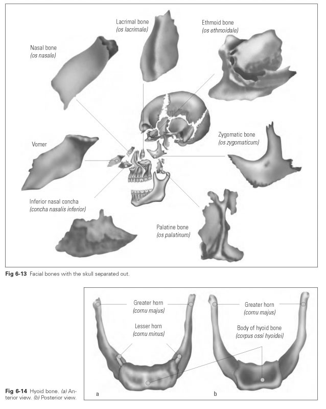
The hyoid bone (os hyoideum) is a small, bracelet-shaped bone that is suspended freely between various muscles (Fig 6-14). In other words, it does not articulate with and is not permanently joined to any other bone. Muscles and ligaments of the floor of the mouth and the throat are fixed to the hyoid. Two processes projecting upward sit on either side of the body of the hy-oid: the large, dorsally placed greater horns (cornu majus) and the small, cartilaginous, centrally placed lesser horns (cornu minus). This is where the stylohyoid ligament attaches, which extends to the styloid process of the temporal bone.