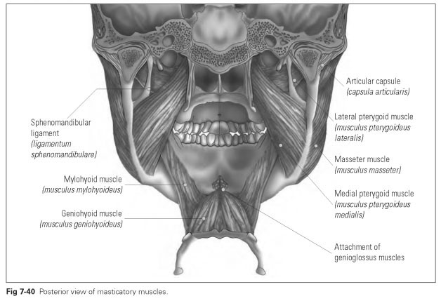
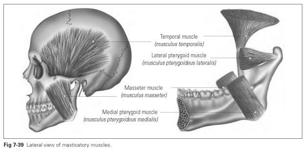
Masticatory Muscles
The temporal muscle (musculus temporalis) is the strongest mouth-closing muscle. Its origin is in the whole temporal fossa, which is almost completely filled. It is made up of a fan-shaped muscle bundle, whose fibers combine to form a very sturdy tendon that runs within the zygoma to the muscular process of the ramus of the mandible. The muscle insertion is actually the whole anterior edge of the ramus of the mandible. The individual bundles of fibers of the fan of muscle partly run in opposite directions.
The fibers running backward draw the mandible backward, while the fibers running vertically work as true mouth-closing muscles; those running obliquely forward are also able to pull the mandible forward. After tooth loss, when this muscle is no longer fully stressed, the muscle mass atrophies and the temporal fossae become sunken. In older people, this reinforces the impression of aging.
The lateral pterygoid muscle (musculus ptery-goideus lateralis) is the muscle that is involved in all movements of the mandible. It comprises two muscle bundles, the upper head of which has its origin on the external surface of the lateral lamina of the pterygoid process of the sphenoid bone (lamina lateralis processus pterygoidei). The lower head of the muscle originates rather deeper at the infratemporal surface of the sphenoid (facies infratemporalis ossis sphenoidalis). The upper muscle bundle runs backward to the capsule of the TMJ and into the articular disc, suggesting that the disc developed from the tendon of this muscle. The lower bundle converges toward the pterygoid fossa of the neck of the mandible below the condyle.
The lateral pterygoid muscle pulls the condyle and the articular disc forward. When the muscles contract on both sides, the mandible moves forward. When it contracts on one side, a lateral movement is performed. The lateral pterygoid muscles are always involved in jaw opening.
The masseter muscle (musculus masseter) has its origin at the zygoma and the zygomatic arch and its insertion at the mandibular angle, externally at the masseteric tuberosities (tuberosita-tes massetericae). The muscle is divided into a sturdy, superficial part (pars superficialis) and a deeper-lying part (pars profunda). The fibers of the superficial part run from the zygoma, coming obliquely downward and backward to the vertical edge of the ramus at the mandibular angle. The fibers of the deeper part run almost perpendicular from the zygoma to the inferior edge of the mandibular body.
The masseter is a powerful mouth-closing muscle that lies along the surface of the side of the face. The two muscle bundles cross over and form a pocket that is open posteriorly. The direction of pull of both bundles produces a rocking movement around the first molar.
The medial pterygoid muscle (musculus ptery-goideus medialis) originates in the pterygoid fossa of the pterygoid process of the sphenoid (fossa pterygoidea) at the internal lamina of this process and runs obliquely downward and backward to the mandibular angle, inward to the pterygoid tuberosities. A few fibers also have their origin at the infratemporal surface of the maxilla. As these sites of origin lie closer together than the sites of
insertion on the mandible, the muscle rather fans out toward the mandibular angle.
The medial pterygoid muscle runs parallel to the masseter and therefore also has the same direction of pull as a mouth-closing muscle. It raises the mandible and, like the superficial part of the masseter, may be involved in protrusive movements.
The masseter and the medial pterygoid muscle form a loop around the mandibular angle in which the mandible lies.The muscle bundle of the masseter is directed upward and outward from the mandibular angle, while that of the medial pterygoid is directed upward and inward. These two pull components—those directed outward and those directed inward —balance each other out during simultaneous contraction, which is why these two muscles can be seen as synergistic (acting together).
During lateral movements, both muscles may also become active. When the jaw is moved to the right, the medial pterygoid muscle and the masseter muscle on the left become active; lateral movement will be supported by the fibers running obliquely forward and upward from the mandibular angle to their origin.
Figures 7-39 and 7-40 illustrate the masticatory muscles from different views.