Molars
The molars (dentes molares) are ideal for the complete crushing and grinding of solid foods. The occlusal surfaces are capable of withstanding the greatest masticatory pressure. In the maxilla, the lingual cusps have an occlusion-fixing function and are referred to as crushing cusps. The buccal cusps have cutting functions and are referred to as shearing cusps. In the mandible, the crushing cusps are buccal and the shearing cusps are lingual.
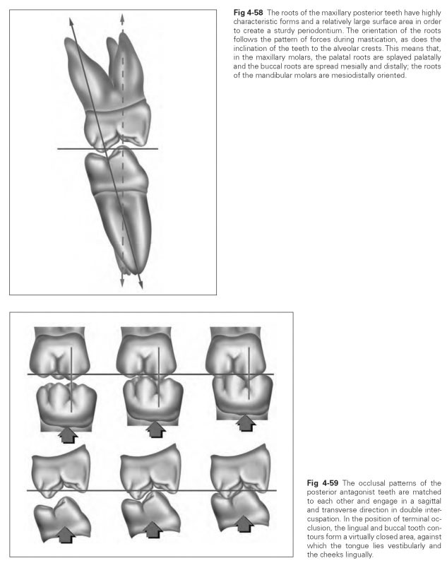
The maxillary posterior teeth show a vestibular inclination. The roots of the maxillary posterior teeth are very characteristically shaped and have a relatively large surface area for a sturdy periodontium. The ratio between the occlusal surface and the surface area of the periodontium is approximately 1:5. At the apices of the roots, there are often small divisions in the root canal, similar to a river delta, which make it difficult to clear the root canal, if necessary. The roots are aligned according to the pattern of forces during mastication, as is the inclination of the teeth to the alveolar crests. The two buccal roots sit almost centrally under the crown, while the distal root is inclined posteriorly and the mesial root anteriorly. This means that central pressures as well as slightly transverse forces that occur within the dental arch can be absorbed. The palatal root is directed palatally and thus absorbs central masticatory pressure and transverse loads.
The mandibular molars have two roots, one distal and one mesial. Both roots have their own canal. These canals can terminate in several outlets at the apex of the root. Development of the pulp horn reflects that of the cusp tips.The prominent roots of the mandibular posterior teeth, like their opposing teeth, have large root surface areas for a robust periodontium. The tooth inclination is not as pronounced on the molars as on the premolars.
Maxillary molars
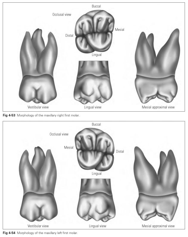
The maxillary first molar (dens molaris medialis) (Figs 4-52 to 4-54) is typical of the maxillary molars. It is the largest of the three large posterior teeth and has all the features characteristic of this form. The occlusal surface contains four cusps, which are separated by developmental grooves.
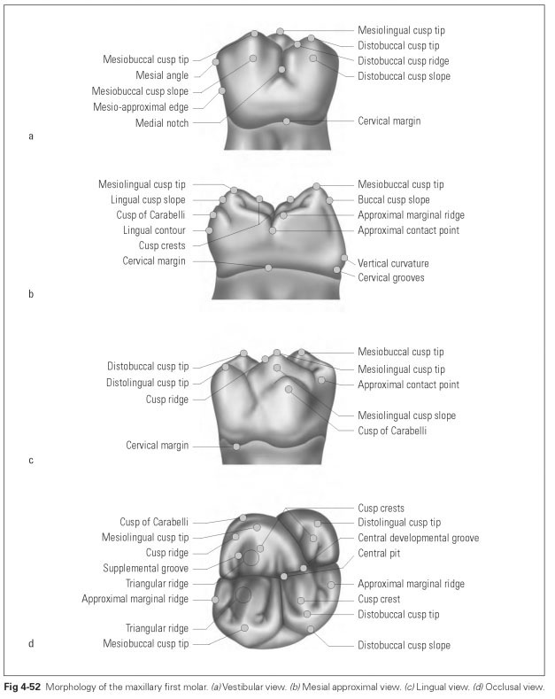
The curvature characteristic clearly stands out. The vestibular and lingual surfaces converge distally to create the typical rhomboid shape of the maxillary first molar.The occlusal surface recedes distally. The maxillary first molars have three roots, two buccal and one palatal.
The average dimensions of the maxillary first molar are:
- Crown width (mesiodistal): 10.5 mm
- Crown depth (buccolingual): 12.0 mm
- Crown length: 7.7 mm
- Total apicocoronal length: 23.5 mm
The maxillary first molar is the first accession-al tooth in the permanent dentition and is usually the first tooth to be lost during exfoliation. It determines the height of the occlusion and the maxillomandibular relationship. This tooth gives the subsequent teeth their positional orientation at exfoliation. During the transitional phase from primary to permanent dentition, it takes on most of the masticatory work. It is ideally suited as an abutment tooth for partial dentures and should be retained as long as possible.
The vestibular (buccal) surface (see Fig 4-52a) gives the impression of being two premolars fused together because it is divided by a distinct longitudinal groove. The mesial and the distal portions of the surface have virtually the same form as a premolar: The occlusal border shows the ridge-shaped cusp form, with the mesial cusp higher and more pronounced than the rounded distal cusp. The medial ridges of the mesial and distal parts of the surface divide each of these into two facets. The mesial part of the surface is more bulging and prominent, while the distal part recedes posteriorly (curvature characteristic). The cervix curves in the middle in an occlusal direction.The cervical grooves are poorly developed.
The approximal surface (see Fig 4-52b) has an almost rectangular shape. The typical vertical curvatures of the buccal and lingual surfaces can be seen.The buccal surface has its greatest curvature cervically, whereas occlusally it has a rather sloping and relatively sharp-edged course up to the cusps.The lingual surface bulges considerably so that the lingual cusps appear to be inclined toward the occlusal surface. The cusp tips on this tooth are also about half the tooth width apart. The mesial approximal surface is much larger (and particularly higher) than the distal; the mesial marginal ridge and hence the contact point are higher.The cervix curves evenly in an occlusal direction.
The lingual surface (see Fig 4-52c) is smaller than the buccal surface, in keeping with the constriction caused by the dental arch. There is also some tapering toward the cervix.The longitudinal groove, which separates the two cusps, is displaced distally because the distopalatal cusp is generally only half the size of the mesial. The mesial cusp is again higher, more angular, and more noticeable. Both cusps, however, bulge inward toward the occlusal surface. The occlusal contour recedes distally.The cervical margin curves occlusally, as on the buccal surface.
The cusp of Carabelli (tuberculum anomale) is an additional, small, low-lying cusp on the mesial part of the lingual surface of the maxillary first molar.
The occlusal surface (see Fig 4-52d) displays typical functional characteristics with four pronounced, differently sized cusps: two buccal shearing cusps and two palatal crushing cusps. Cusps in order of decreasing size are: mesiolin-gual, mesiobuccal, distobuccal, and distolingual.
The structure of the individual cusps reflects the described features.The buccal cusp ridge and crests are angular, whereas the lingual cusps appear rounded.
The developmental grooves form small pits at their crossover points. Where the central developmental groove comes into contact with the buccal groove, the compact central fossa is formed. The supplemental grooves at the marginal ridges also form pronounced pits at the branching points with the central groove. The shape of the grooves produces a skewed, rather distorted H. The marginal ridges in the approximal area are noticeable, while the mesial approximal edge is rather higher, almost straight, but the distal edge is curved outward. The marginal ridges form the contact points in the transitional area with the ap-proximal surfaces and form the approximal depressions with the adjacent teeth.
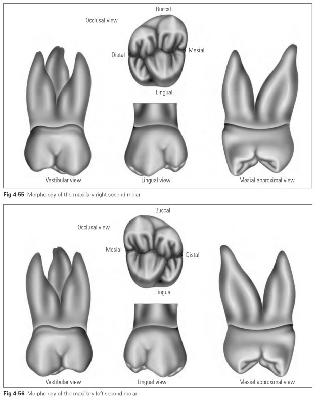
The maxillary second molar (dens molaris laterali) (Figs 4-55 and 4-56) has the same form as the first molar, with the only difference being that its lingual surface is less developed. The outline of the crown is often modified so that the rhomboid shape appears more acutely angled and the whole crown is far smaller than that of the maxillary first molar.
The cusp of Carabelli is absent, and the distolingual cusp is smaller, sometimes shrunken to a marginal ridge so that the occlusal surface has only three cusps. The three tooth roots are often fused.
The average dimensions of the maxillary second molar are:
- Crown width (mesiodistal): 9.8 mm
- Crown depth (buccolingual): 11.5 mm
- Crown length: 7.7 mm
- Total apicocoronal length: 21.1 mm
The vestibular (buccal) surface is divided by a distinct longitudinal depression as in the maxillary first molar. The mesial cusp is higher and more pronounced than the rounded distal cusp, which recedes sharply. The mesial aspect of the surface is much more convex and prominent than the part that recedes distally. The cervical line bends occlusally in the middle. Cervical grooves are only poorly developed.
The lingual surface is much smaller than the buccal surface, tapering sharply to the cervix.The distolingual cusp may be rudimentary so that the occlusal contour sharply recedes distally. The mesial cusp is again higher, more sharp-edged, and more developed. There is rarely a cusp of Cara-belli present.
The approximal surface has an almost rectangular shape. The typical vertical curvatures of the buccal and lingual surfaces can be seen. The mesial approximal surface is also much larger here than the distal surface, with the mesial contact point located much higher.The cervical line bends occlusally.
The occlusal surface also displays typical functional characteristics, usually with four differently sized cusps: two buccal shearing cusps, one lingual crushing cusp, and one distolingual cusp shortened at the marginal ridge. The central developmental groove with the main buccal groove forms the central pit. The mesial approximal marginal ridge is more pronounced and higher, almost straight, while the distal margin curves outward again.
The maxillary third molar (dens serotinus) is the most variable of all teeth: from a four-cusp form to a small peg tooth.The tooth can have three roots, but the peg forms often have only one root. Certain forms also have several root apices.The third molar appears to be in the process of regression, ie, the dentition is being reduced, with the third molar often not developing.
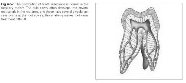
Details of the pulp cavity, roots, and occlusion of the maxillary posterior teeth are presented in Figs 4-57 to 4-59.