Immune defense
Dentin-cellulose complex is uniquely organized for offset microbial threats from caries, as well as other violations external hard fabrics of a tooth in the oral environment. While permeable for bacterial elements by virtue of the dentinal tubules, dentin, however, performs an important function filter significance pulp immune protection. First of all, two mechanisms to account for this effect: (i) peripherally directional flow of dentinal fluid, and (ii) the absorption of bacteria and bacterial macromolecules for the internal walls of tubules (63). Thus, dentin can moderate the impact of harmful elements cellulose, allowing it to adapt and to organize an effective immune response.
The components making up the innate immune protection pulp include resident tissue cells, viz. odontoblasts and immune cells, nerves and blood vessels (Fig. 2.12). Basal configure immune cells is limited antigen-presenting cells (APC), macrophages and T-lymphocytes (T-cells) memory type (23). Neither B-cells, neither fat cells, are residents of the normal pulp and will not appear in the fabric, if there is an inflammatory events.

The APCS ordinary cellulose are of two types.
One has a pronounced dendritic configuration and belongs to the family of dendritic cells (DC). Another monocyte/macrophage lineage. As essentially carry class II MHC molecules on the surface of cells. This molecule is present in all cells capable of antigen presentation of the gene product of the major histocompatibility complex (MHC), and acts as a stimulating molecules in T-cell activation in local and regional lymph nodes.
DCs in the pulp strategic positions in the periphery of the tissue where the foreign antigens are likely to enter (Fig. 2.12 and 2.13). They compete for the available space with odontoblasts and make contact with these cells through their cytoplasmic processes (52). The pulp of a tooth DCs are also in close proximity with elements of the nervous tissue and blood vessels in paraodonto-cynical region, offering interactive potentials (53, 54).

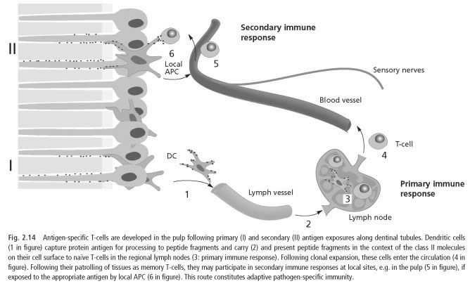
The main function of DCs to alert the immune system to effectively subsequent elimination, and not directly combat the invading microorganisms. In peripheral sites as mass, they occur in an immature state. Maturation become a professional APCS (capable of activation of T cells that were not exposed to the antigen in front of the so-called " naive T-cells) comes from the capture incoming foreign antigens. When the impact they usually begin to migrate to the regional lymph nodes. Once there, fragments of antigen is bound to a class II MHC molecules will show the corresponding naive T-cells, which are activated in the primary immune response (Fig. 2.14). DCs are especially effective in microbial sensing, capture and processing of foreign antigens and, thus, the key initiator of the adaptive immune response.
Class II molecules-expressing macrophages are distributed in a noticeably higher number in the pulp and appears to form a dense network together with the tooth pulp DCs (33, 34). As in other connective tissue macrophages in the pulp heterogeneous in terms of the phenotype and function. Although there are those who serve in the local antigen presentation, there is a large population of rooms class II molecules-expressing, a resident macrophages (histiocytes), primarily located perivascularly with the main functions of phagocytosis.
 ..
..