Alveolar Crestal Bone
1.
Alveolar bone is that part of the bone that support the teeth.
2. The surface of the bone combs, smooth, covered with a thin layer of bark (dense, solid bone, which can be considered as a thin white line on the x-ray.
3. The most important radiographic features of the alveolar ridge, is that it forms a smooth undamaged surface between adjacent teeth with only the Width of the periodontal ligament space, separating it from the adjacent surface of the root.
a. The crest of interdental partitions between the incisors - thin and sharp.
B. The crest of interdental partitions between the back of the teeth is round or flat (Fig. 20-4).
E. Lamina Is " Stupid"
1. Alveolar bone is actually a thin layer of dense bone tissue that lines the normal tooth socket. In x-rays, alveolar ridge proper defined as lamina-dur. On the x-ray lamina dura looks like a solid white (opaque) line around the tooth root (Fig. 20-5).
2. On the x-ray lamina dura continuously with the cortical bone comb from interdental partitions.
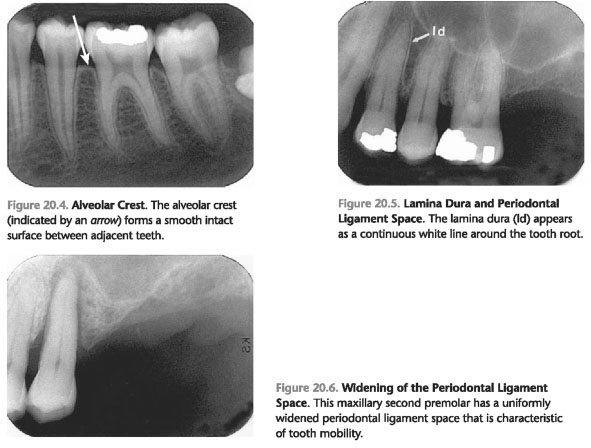
F. Periodontal Ligament Space
1. The space between the root of the tooth and lamina dura socket is populated connective tissues of parodentium. Periodontal ligament tissue functions as an attachment teeth for the record dura socket.
2. Periodontal ligament tissue does not resist the penetration of x-rays and, therefore, appears on the x-ray as thin radiolucent black line around the tooth root (Fig. 20-5).
3. In most cases, the expansion of the periodontal ligament space (PDLS) on the x-ray indicates, tooth mobility (Fig. 20-6).
 ..
..