Muscle as a Motor Unit
Muscle contractions are triggered by nerve impulses. In the case of voluntary movements, the nerve impulses are transmitted by motor nerve pathways from the cerebral cortex via the spinal cord to the motor end plates on the individual muscle fibers. The number of muscle fibers in a muscle may be as high as two million. Each muscle fiber can be stimulated independently and contract separately from the other fibers. All the muscle fibers can be stimulated at once by the nervous system, or only some of them can be stimulated and a movement can be performed with graduated expenditure of energy. Destruction of nerve conduction results in paralysis of the muscle and inactivity atrophy; ie, the muscle shrinks or atrophies. Muscle mass will increase if put under heavy strain.
Skeletal muscles have a uniform structure: The ends of a muscle are referred to as the insertion and the origin (sites of attachment), while the actual muscle mass is known as the belly (venter). The origin (caput) denotes the point where the muscle attaches to the passive locomotor system. Contraction of the muscle mass pulls the point of insertion and point of origin closer together so that the bones are moved, mostly via joints. However, there are also muscles in the locomotor system without a specific origin and insertion, eg, certain facial expression muscles. As a rule, the skeletal muscles always have corresponding antagonists that accomplish the same movement but in reverse. This is not actual antagonism but a functional interaction, even where the pull is in opposite directions, because this is the only way that coordinated movements can take place.
Muscle tone refers to the resting tension when a muscle is in its relaxed state. The tone ensures that certain permanent postures can be assumed, eg, that of the trunk when the body is erect. The tone of the masticatory musculature holds the mandible in the physiologic rest position.
Contraction of a muscle takes place through a very rapid succession of individual shortenings of the muscle fibers. To maintain continuous shortening, the nervous stimulation has to activate the muscle periodically; in other words, the muscle has to be switched on and off. In terms of frequency, the muscle starts to vibrate and thus produces a muscle tone of roughly 30 Hz.
Muscle power can be determined in relation to the cross-sectional area of the muscle: A muscle with a cross-sectional area of 1 cm2 produces an absolute muscular power of about 100 N. Certain auxiliary organs, such as nerves and blood vessels, tendons and tendon sheaths, and bursae and fasciae belong to muscles.
Tendons are made up of white, shiny, parallel fibers of collagenous connective tissue with high tensile strength (50 N/mm2). They emerge from the combination of sheaths of individual muscle fibers and generally attach in the periosteum of the bones. However, direct insertion of a tendon into the bone may also occur.
Tendon sheaths affix tendons to the bone or joints to safeguard the functioning of the tendons themselves. The tendon sheath secretes mucus-similar to synovial fluid in the joints—so that the tendon is able to glide back and forth smoothly.
Bursae are sacs or saclike cavities found around joints that act like a cushion of water; ie, they hold pressure away from the bone to prevent bone destruction. Bursae are found wherever tendons pass close by the bone, especially at the joints.
Fasciae are sheaths of connective tissue around individual muscles or muscle groups. The fasciae form guiding sheaths and hold the muscles in the correct position in their relaxed state. When they are particularly sturdy and tendonlike, fasciae may also form points of origin and insertion for muscle fibers.
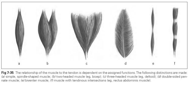
Muscles vary in shape; they can be short, long, wide, or flat (Fig 7-35). Skeletal muscles are classified according to their tendinous parts:
- Spindle-shaped muscles with a tendon of origin and insertion on a spindle-shaped belly; simple, two-headed (bicipital), three-headed (tricipital), or multitailed muscles are possible (see Figs 7-35a to 7-35c)
- Pennate (feathery) muscles with a central tendon from which the muscle fibers emanate on one or both sides (see Fig 7-35d)
- Fan-shaped muscles are multiserrated or multidivided muscles
- Muscles with tendinous intersections, eg, biventer muscles (mylohyoid muscle) (see Figs 7-35e and 7-35f)
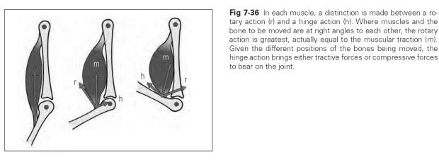
In each muscle, a distinction is made between a rotary action and a hinge action. Figure 7-36 illustrates this distinction and what kind of forces each action bears on the joint.