Perioral Muscles of Facial Expression
The muscles of facial expression (also known as "mimic muscles") do not run from one bone over a joint to another bone but often only attach to the skin, without an intermediate tendon. The facial expression muscles move the facial skin and give the face its expressiveness. The numerous furrows, dimples, and creases that are produced by these muscles are an expression of a person's mood. The facial expression muscles are mainly arranged around the orifices of the mouth, nose, and eyes, as well as the ears, because the position and shape of these openings in the face determine the particular expressive quality of the face.
The perioral muscles of facial expression are the muscles of the mouth and cheek area that belong to what are known as the accessory masticatory muscles and play an important role in eating (Fig 7-43). They form the bulk of the muscular basis of the external wall of the vestibule of the mouth (cheeks and lips), while their sites of origin influence the position of denture borders (Figs 7-44 to 7-46). Furthermore, the movement of the facial expression muscles can jeopardize the retention of a complete denture but also stabilize it. To make use of this stabilizing force, the denture base must make allowance for the position and path of the muscles at rest and during activity.

In advanced age, the muscles of facial expression atrophy so that they lie more or less loosely against the teeth. In the area of the posterior teeth, there is a risk that patients will bite their cheek. This can be counteracted when shaping the denture borders, while supporting the stability of the denture at the same time.
The external surfaces of the denture body are shaped so that they grip the muscles. In the anterior area, lip shields for the orbicular muscle of the mouth are prepared; the border of the denture should be chamfered cervically above the vestibular fornix so that the orbicular muscle is firmly engaged to stabilize the denture.
In the posterior area, buccinator supports are created and the muscular tracts are redrawn on the ligaments of the cheek. The denture border is shortened in keeping with the oblique line (buccinator attachment) and widened horizontally into the cheek. As a result, the cheek lies on the border and presses the denture onto the jaw, while at the same time the cheek is pushed away from the teeth so the patient will not bite it.
The path of the muscular tracts, starting from the modiolus in the angle of the mouth, can be utilized when shaping the denture body to make it grip the muscles (Fig 7-47). The denture acrylic resin must not be shaped too thickly in the labial vestibule because this will widen the vestibular fornix as if the patient had a roll of cotton wool there, drawing the red of the lips inward (see Fig 7-45).
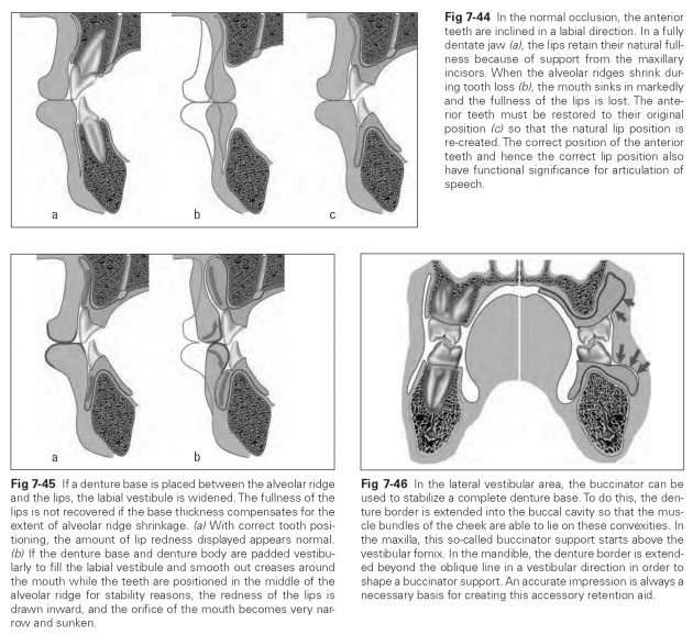
The anterior teeth should stand within the vertical anterior dental arch so that the incisors fill out the upper lip with their labial surface and can support the lower lip with their incisal edges.
In the maxilla, the buccal vestibular space should be shaped thinly because the masseter presses the buccinator into the vestibule here. One way of gaining an overview of the large number of muscles of facial expression is to start from a distinctive point and look at the muscles grouped around that point.
In this model, the corner of the mouth (labial commissure) should be chosen as the orientation point for the following reasons: Starting from the corner of the mouth, antagonistic muscle groups can be described, which provides a certain overview. Around the corner of the mouth, six essential, partly antagonistic muscles of the perioral region come together in the modiolus.
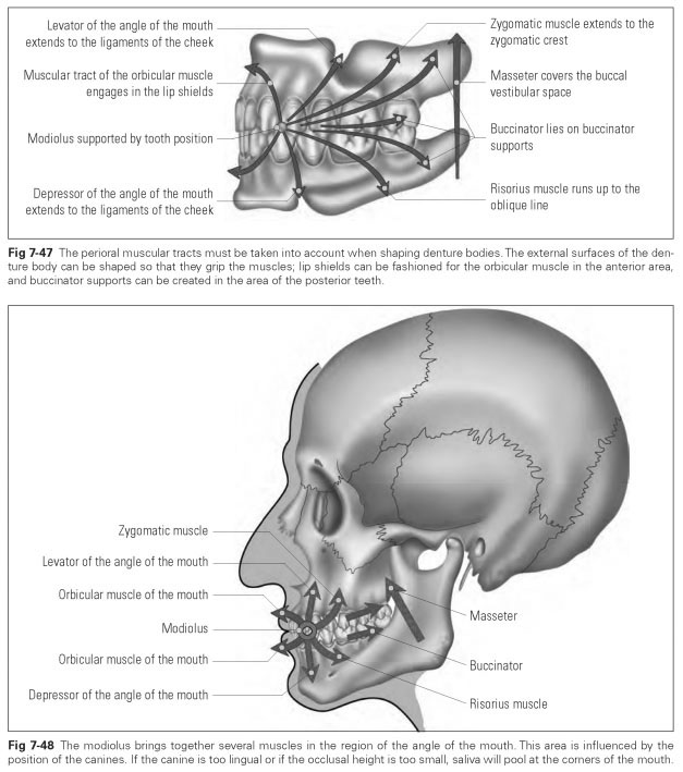
The modiolus is a small aponeurosis (a tendinous plate) into which most of the muscles of facial expression attach (Fig 7-48). Consequently, the corner of the mouth is particularly mobile, which should be taken into account when shaping the denture. As a result of the position of the mandibular anterior teeth, the modiolus may help to stabilize a mandibular denture if it lies higher than the mandibular anterior teeth and is able to press onto the incisal edges. Therefore, the mandibular incisors should never stand higher than the lip closure line.
The arch width of the mandibular anterior teeth must also be kept in correct proportion because, if it is too wide in the canine area, the denture may be lifted by muscular activity (eg, that of the levator muscle of the angle of the mouth).