Primary Teeth
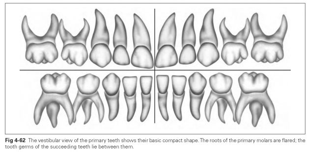
The teeth of the primary dentition are known as deciduous (dentes decidui) or primary teeth as well as the mixed or temporary dentition, and they form the masticatory apparatus up to the sixth year of life. The primary dentition is made up of 20 teeth: eight incisors, four canines, and eight primary molars. The forms of these teeth match those of the permanent dentition, but there are no premolars (Fig 4-62).
The primary teeth that stand in place of the premolars bear a closer resemblance to the molars of the permanent dentition, which is why they are known as primary molars. The primary teeth are smaller and more delicate than the permanent teeth (2 to 4 mm smaller on average). They look bluish-white, in contrast to the yellowish color of the permanent teeth, because they contain less calcium.
One striking feature of the primary teeth is a thickening of the enamel margin cervically, so that they bulge outward over the gingival margin. The size ratio is an equally reliable characteristic: The primary teeth are far smaller but relatively broader than the permanent teeth in relation to their length; seen from the front, the primary dentition appears shorter than the permanent dentition. It should be noted that variations in the form of the primary teeth are very rare.
The anterior teeth of the primary dentition largely resemble the teeth with the same name in the permanent dentition.The incisors and canines are smoother than those in the permanent dentition, with less pronounced ridges. However, the central ridge is noticeably well developed labially, both in the maxilla and in the mandible. All the features, especially the angle characteristics, are strongly developed. The lingual surfaces in turn are less pronounced; ie, the dental tubercle is only slightly developed.
The primary maxillary first molar resembles a permanent premolar, but its pronounced rhomboid shape is noticeable.This tooth may also have a second cusp on the lingual half. The distinctive fissure separates the buccal and lingual cusps.
The primary maxillary second molar has some similarities with a permanent maxillary molar, but the rhomboid shape is predominant, while the mesiobuccal cusp overhangs a great deal buccally. The primary mandibular first molar bears no resemblance to the permanent premolar destined to replace it. It usually has four cusps on the occlusal surface, with the mesial cusps dominant. The cusp tips are sharp and pointed, as they are on all the primary molars. Ridge formation on the occlusal surfaces is prominent.
Among all the primary molars, the primary mandibular second molar bears the strongest resemblance to its succeeding molar, except that the middle buccal cusp protrudes slightly buccally and the cusps are more pointed and more prominent overall.
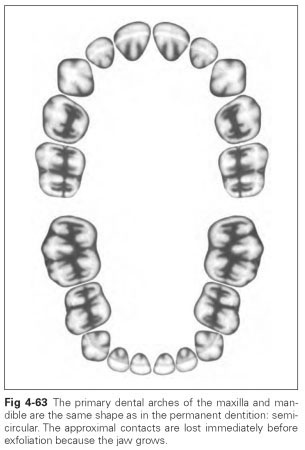
The dental arch form of the primary dentition is similar to that of the permanent dentition (Fig 4-63). The approximal tooth contacts are lost immediately before exfoliation of the teeth in approximately the sixth year of life because the jaws become larger.
The roots of the primary molars are widely flared because the tooth germs of the eventual premolars have to find space between them (see Fig 4-62). The pulp cavity and root canal of the primary teeth are spacious, and the walls of the teeth are correspondingly thinner. The roots of the primary incisors in the maxilla and in the mandible are quite flattened, whereas the roots of the primary canines are more evenly rounded. At exfoliation, the roots of the primary teeth are resorbed (resorb = to suck back) as a result of pressure from the growth of the permanent teeth.
The primary teeth are extremely susceptible to caries, which often leads to premature tooth loss or, in extreme cases, damage to the succeeding tooth. Because the primary teeth act as space maintainers for the permanent teeth, they should be retained for a long time. Any premature loss can influence the growth of the jaw, resulting in a lack of space for the permanent teeth and hence positional anomalies.