Cranial Anatomy
The bony skull (cranium) forms the basic bony structure (skeleton) of the head. It is balanced on the first cervical vertebra or the spinal column. It is the point of origin and attachment of many muscles, which in turn partly defines its shape. The shape of the skull is mainly determined by its contents. For instance, there is a connection between the brain and the structure that houses the brain; if the brain grows excessively, the cranium surrounding it becomes enlarged. Equally, premature ossification of the cranium leads to malformations of the brain. A similar relationship exists in the facial part of the skull, where special functions such as that of the masticatory system enforce a specific shape on the jaws, the temporomandibular joints (TMJs), and the teeth.
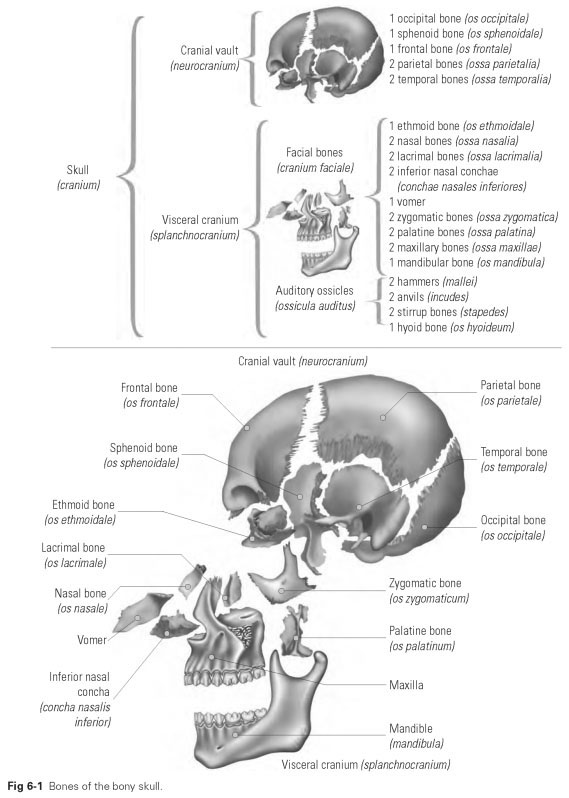
The cranium is made up of 29 cranial bones, which are fitted together like a mosaic to make a roughly spherical shape (Fig 6-1). The individual bones are fused together by sutures, cartilage, and bony connections.The exceptions are the mandible, which is fixed to the skull by two joints, and the hyoid bone, which does not articulate with and is not firmly fixed to any bone in the body but is suspended freely between muscular tracts.
If the skull is divided by a circular, horizontal cut at its widest circumference, the result is the high-vaulted roof of the skull (calvaria) and the base of the skull. On the inside of the calvaria as well as the cranial sutures, there are impressions, fossae, and depressions that are produced by blood vessels and by the surface of the brain.
The skull cap (calvaria) forms the superior, closed, spherical shell of the skull, which is formed by the parietal bones, the frontal bone, and the occipital bone. The calvaria is made up of lamellar bony plates and extends from the orbital margins to the midpart of the occipital bone and at the sides to the temporal bones.
From the cut surface of the skull cap, the structure of a typical bony plate is visible, with an outer layer (lamina externa) and an inner layer (lamina interna); between the two compact laminae lies spongy or cancellous material of varying thickness, which contains the bone marrow and numerous blood vessels. Very thin-walled veins run from the bone marrow in and out through special bone canals. The outer surface of the skull cap is covered by a relatively thick layer of periosteum, the pericranium. On the interior surface, the periosteum is represented by the dura mater of the brain, which adheres to the bone.
The skull, as the top of the body, encases the brain and the sensory organs and contains openings for the digestive tract and the airways. The different functions of the skull determine the differentiated form of its two different areas: the cranial vault and the facial bones.
This division into the cranial vault and the visceral cranium is most marked when viewed from the side. The border between the two parts runs roughly around the root of the nose, over the supraorbital margin, and out to the outer auditory orifices.
The cranial vault (neurocranium), formed by the temporal bones, the sphenoid bone, the frontal bone, the parietal bones, and the occipital bone, is virtually round and relatively smooth. The external auditory canal plus the zygomatic bone (also running from the temporal bone), as well as a prominent muscle attachment to the temporal bone (the mastoid process), are the most striking features on the lateral surface of the skull.
The facial bones, or the visceral cranium (splanchnocranium; Greek, splanchnos; Latin, viscus = viscera), are mainly formed by the dominant maxilla and mandible. They also include the zygomatic bones, nasal bones, lacrimal bones, ethmoid bones, inferior nasal conchae, vomer, palatine bones, hyoid bone, and the auditory ossicles (hammer, anvil, and stirrup).The zygomatic bone forms the lateral orbital margin as well as the attachment to the zygomatic arch.The maxilla forms the inferior edge of the orbit and the lateral borders of the nasal cavity.The frontal bone forms the bulging superior orbital margin.
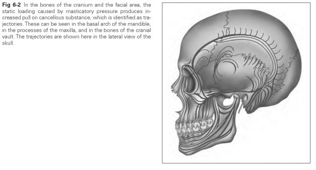
Pronounced cancellous tracts, known as trajectories, can be seen in the facial bones and the bones of the cranial vault. These develop from the static loading caused by masticatory pressure (Fig 6-2).They can be seen in the basal arch of the mandible, in the processes of the maxilla, and in the bones of the cranial vault.