Mandibular premolars
The main difference between the mandibular and maxillary premolars is that the mandibular ones have an almost circular crown outline. Furthermore, they are always single-rooted. Unlike the maxillary premolars, the mandibular first and second premolars differ considerably from each other.
The mandibular first premolar (Figs 4-46 and 4-47) is similar to a mandibular canine, except that the dental tubercle is developed into a very small, independent cusp. As the secondary antagonist of the maxillary canine, the buccal cusp of the mandibular first premolar is most strongly developed. Particularly tough food is often chewed by the maxillary canines and their opposing teeth (the mandibular canine and first premolar).
The average dimensions of the mandibular first premolar are:
- Crown width (mesiodistal): 6.8 mm
- Crown depth (buccolingual): 6.8 mm
- Crown length: 8.4 mm
- Total apicocoronal length: 22.0 mm
The collum angle or crown-root angulation (Fig 4-48a) on the mandibular premolars is an angle between the tooth axis and the crown axis. If an axis is drawn through the tooth from the root apex to the tip of the buccal cusp, and another is drawn from the base of the crown to the central developmental groove, there is a striking difference between the paths of these axes.The bend in the root in the crown-root angulation means that the crushing buccal cusp sits centrally, ie, in a statically favorable position over the base of the crown, and strain is placed axially on the periodontium.
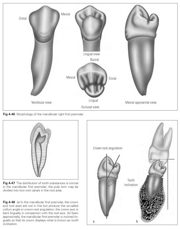
The tooth inclination (Fig 4-48b) refers to the distinct lingual incline of the mandibular posterior teeth.The tooth inclination is most pronounced on the first premolar, and the degree of inclination declines distally on the posterior teeth.
The vestibular (buccal) surface of the first premolar is very similar to that of the mandibular ca-nine.The first premolar is only slightly more compact, and the contact areas may be rather tapered. Overall the surface is highly convex. The ridgeshaped cusp has a rounded tip, while the mesial cusp ridge is shorter than the distal (angle characteristic). One prominent central ridge divides the buccal surface again into two unequally sized facets with vertical depressions. The mesial contact area lies higher than the distal. In the cervical third, a short transverse and longitudinal convexity can be seen with poorly developed cervical grooves. The arched line of the cervix converges with concave, curved approximal margins. The curvature characteristic, like the angle characteristic, is well developed.
The lingual surface is very small and narrow and shows the very slightly developed lingual cusp. It tapers more cervically than buccally. The buccal cusp can be seen from the lingual aspect; only the central developmental groove is concealed by the small lingual cusp.This cusp has no opposing contact. As a result of the tooth inclination, the lingual cusp greatly overhangs the cervix; however, it is highly concave in the incisal third, so that a pronounced vertical curvature is visible.
The approximal surface reveals both the large buccal and the small lingual cusps. The lingual bend in the crown axis corresponding to the tooth inclination is most clearly visible approximally, as is the prominent longitudinal convexity—buccally in the cervical area and lingually in the occlusal. The approximal surfaces are prominent at the contact area and concave cervically.
The occlusal surface shows the circular outline of the crown. The lingual cusp is much smaller than the buccal and also more truncated. The occlusal surface is therefore markedly inclined toward the floor of the mouth. A sturdy cusp crest runs lingually from the buccal cusp; as a result, the central developmental groove is sometimes interrupted. The central developmental groove is markedly displaced lingually.
The approximal marginal ridges are sturdy and recede in a lingual direction. This produces two distinct fossae, with the mesial one being more superior. The distal marginal ridge also lies more inferiorly. There are three variations on the arrangement of the lingual cusp:
- There is a very regular arrangement of the lingual cusp, where the line connecting the two cusps divides the tooth symmetrically.
- The lingual cusp is small and rudimentary like a tubercle; there is only a suggestion of a central developmental groove.
- The lingual cusp is displaced distally so that the crown takes on a triangular shape with the buccal cusp tip displaced mesially.
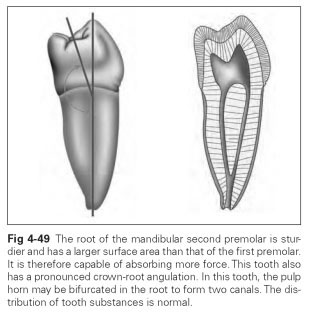
The mandibular second premolar is larger than the first premolar, but the resemblance is not as strong as that seen between the first and second premolar in the maxilla. The occlusal surface is more horizontal; ie, there is only a slight difference in height between buccal and lingual cusps. The tooth assumes two common forms. One has two cusps, and the other has three cusps, with one buccal and two lingual cusps. Very rarely there may be a four-cusp type with one buccal and three lingual cusps. The root of this tooth is roundish and, consistent with the stronger development of the second premolar, is longer and thicker than the root of the first premolar.The root is only bifurcated in rare cases.This tooth is capable of absorbing considerable masticatory forces. The pulp horn undergoes lobelike widening in correspondence with the cusps (Fig 4-49).
The average dimensions of the mandibular second premolar are:
- Crown width (mesiodistal): 7.5 mm
- Crown depth (buccolingual): 9.0 mm
- Crown length: 8.5 mm
- Total apicocoronal length: 24.0 mm
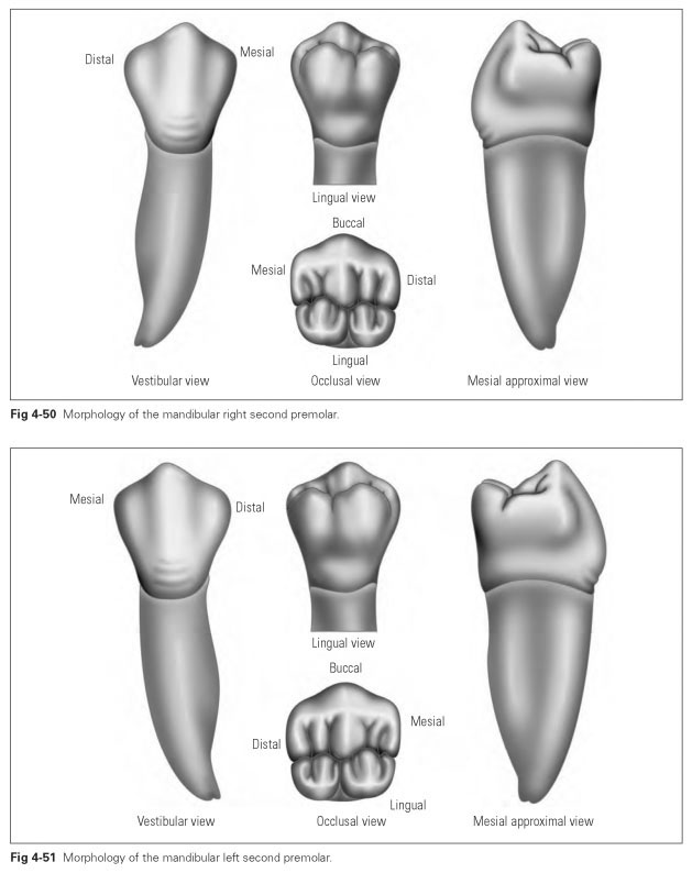
The mandibular second premolar (Figs 4-50 and 4-51) is a tooth with a true masticatory surface in form and function. It also displays tooth inclination and crown-root angulation, although these are less pronounced than with the first premolar. As a result, the tooth makes full occlusal contact with all of its cusps.
The vestibular (buccal) surface resembles a compact, broad canine, with a ridge-shaped cusp ridge and rounded tip. The angle characteristic is pronounced so that the mesial angle lies slightly higher than the distal, as do the contact points. The formation of ridges and depressions is normal, and a curvature characteristic is present.The approximal margins are indented and taper down to the curved cervical margin.
The lingual surface is narrower and slightly shorter and has a pronounced transverse convexity. In the three-cusp type, the two lingual cusps are recognizable and make the surface appear divided. It is noticeable that the distolingual cusp is smaller and lower than the mesial. The surface also appears to overhang in the cervical area because of the tooth inclination and the strong vertical convexity. The buccal cusp can be seen in the lingual view because it rises above the lingual cusps.
The occlusal surface has all the features of a masticatory surface: cusps, cusp crests and ridges, marginal ridges, and grooves. The two-cusp type resembles the form of the maxillary second premolar. In the three-cusp type, the buccal cusp is the largest, while the linguodistal is the smallest. The three cusps are formed by a large main developmental groove that diverges at a right angle from the central developmental groove: This main groove often originates in the middle or slightly more distally. As a result, the groove formation appears to create a Y shape, which divides the three cusps. A third, very rare form is the four-cusp occlusal surface with three lingual cusps.
The approximal surface shows the crown-root angulation, ie, the lingual incline of the crown. In this view, the tooth inclination of the second premolar is also identifiable, but it is less developed than on the mesial neighbor. The vertical curvatures of the buccal and lingual surfaces can also be seen. The lingual contour overhangs in the occlusal area. The occlusal surface is only slightly tilted lingually. The buccal part of the occlusal surface is wider, so that the central developmental groove is displaced slightly lingually. The ap-proximal marginal ridge contains the contact point and tapers so that the approximal surface is concavely indented.