Maxilla
The maxilla is a paired bone; ie, there are two maxillary bones that are joined by a bone suture. However, the individual maxillary bones also have a paired arrangement. Thus, it is still possible to detect the development from one maxillary process and the frontal process, giving rise to the unified maxillary bone for each half of the face.
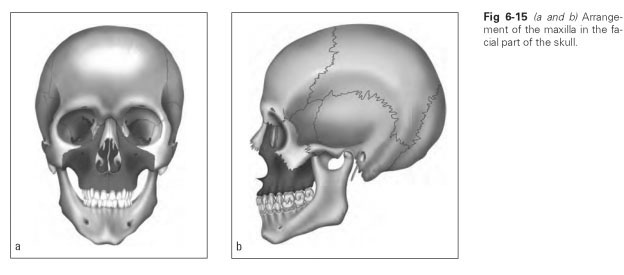
The maxilla, like the mandible, is described as one bone because functionally it represents a single structure. It lies centrally in the facial or visceral cranium and thus forms the bony foundation of the face (Fig 6-15); the shape and size of the face are determined by the dimensions of the maxilla. A compact body of the maxilla, from which the four maxillary processes project, can be identified in each maxillary bone. The walls of the orbit, the nasal cavity, and the palatine vault are formed by the maxilla. It holds the maxillary dentition and transfers masticatory pressure via the processes to the cranial vault.
The body of the maxilla (corpus maxillae) is made up of thin bony plates and contains the maxillary sinus (sinus maxillaris). Four surfaces can be identified on the three-sided body resembling a truncated pyramid: the facial surface, the nasal surface, the orbital surface, and the infratemporal surface.
The facial surface (facies anterior) is the anterior surface of the body of the maxilla, which is bordered superiorly by the infraorbital margin (margo infraorbitalis) and posterolaterally is divided from the infratemporal surface by the in-frazygomatic crest (crista infrazygomatic). A few millimeters below the infraorbital margin lies the infraorbital foramen (foramen infraorbitale), which is the exit for the infraorbital canal (cana-lis infraorbitale) that emerges from the orbit. The canine fossa (fossa canina) is a flat depression located roughly at the root apex of the canine, below the infraorbital foramen and the root of the zygomatic process. It is named after the canine tooth and is the origin of a mimic muscle (muscle of facial expression).
The infratemporal surface (facies infratempo-ralis) is demarcated from the facial surface by the infrazygomatic crest. The protuberance-like bulging of the maxillary tuberosity (tuber maxillae) at the inferior edge of the surface is striking; its rough areas border the palatine bone and the pterygoid processes. Toward the cheek are located the alveolar foramina (foramina alveolaria), tiny holes that are openings for parts of the maxillary branch of the trigeminal nerve for the dental nerves of the molars. A groove running from a posterior-superior to an inferior-anterior position over the tuberosity toward the palate, together with the sulcus of the palatine bone (sulcus ptery-gopalatinus), form the pterygopalatine canal (canalis pterygopalatinus canalis; in the palatine area, also known as the greater palatine sulcus, ie, canalis palatinus major).
The orbital surface (facies orbitalis) forms the largest part of the orbital floor. A groove runs from the posterior orbital margin and anteriorly becomes the infraorbital canal. It is the infraorbital sulcus (sulcus infraorbitalis), the course of which is followed by the infraorbital nerve, part of the maxillary branch of the trigeminal nerve. In the bottom of the sulcus and the canal there are tiny holes through which nerves from the maxillary anterior teeth pass.
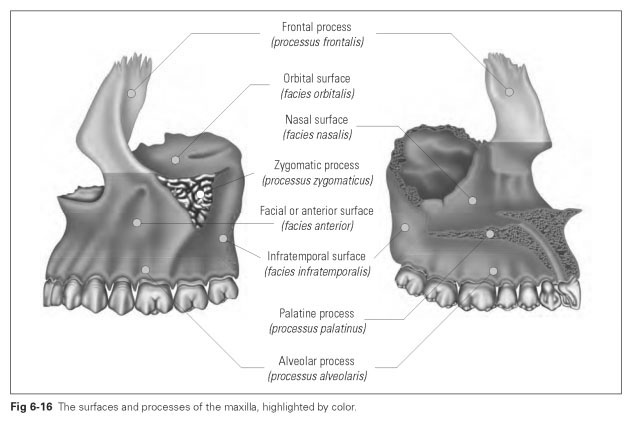
The nasal surface (facies nasalis) forms the inner surface of the corpus and the lateral wall of the nasal cavity. This surface contains a long, narrow, five-sided window known as the maxillary hiatus (hiatus maxillaris), which marks the entrance to the maxillary sinus. This maxillary hiatus is covered posteriorly by the perpendicular plate of the palatine bone, inferiorly by the inferior nasal concha, and superiorly by the ethmoidal labyrinth. There is a prominent ridge to which the inferior nasal concha attaches (crista conchalis). The lacrimal sulcus (sulcus lacrimalis) becomes the lacrimal canal or duct where it is covered by the lacrimal bone.
The maxillary sinus (sinus maxillaris) is a cavity in the corpus of the maxilla that is lined with mucous membrane and filled with air. It is the largest paranasal sinus and is connected to the frontal sinus via the other paranasal sinuses.The apices of the roots of the molars and possibly the premolars project into the sinus and are generally covered with thin bony plates. Sometimes the bony covering over a root apex may be missing so that the root protrudes freely into the sinus.
Figure 6-16 illustrates the surfaces and processes of the maxilla.