Maxillary Processes
The frontal process (processus frontalis) rises vertically between the lacrimal bone and the nasal bone up to the frontal bone. It forms a closed bony connection with the cranial area so that masticatory forces can be transferred to the cranium while bypassing the nasal and orbital cavities.
The zygomatic process (processus zygomaticus) is a short process with a triangular cross section that runs along the side of the orbital surface to the zygomatic bone. A convexity of the maxillary sinus may be found inside the process. This process also transfers masticatory pressure vertically to the cranium, while bypassing the eye socket, but also laterally via the zygomatic bone to the temporal bone.The infrazygomatic crest (crista in-frazygomatica) runs caudally from the zygomatic process to the alveolar jugum (jugum alveolare) of the distobuccal root of the maxillary first molar.
The alveolar process (processus alveolaris) is directed vertically and downward and, with the process on the opposite side, forms the alveolar ridge. It bears the teeth, and its shape is dictated by the dental arch, which is why it is also known as the dental process. This maxillary process is strongly functionally designed and is resorbed following tooth loss.This involves irreversible, re-sorptive disuse atrophy, in which the tissue is broken down by osteoclasts. As a result, the alveolar ridge shrinks in the direction of inclination. All of the other processes are also subject to this process of disuse atrophy, but their shape does not alter as markedly as that of the alveolar process.
At the wide inferior edge of the alveolar process lie the dental alveoli or tooth sockets (alveoli dentales).There are eight tooth sockets, which are separated by interdental or interalveolar septa (septa interdentale, septum interalveolare). These thin sheets of bone can also emerge between individual root tips in the case of multirooted teeth and then form interradicular or alveolar septa (septa interradicularia or septum alveolare). In keeping with the shape of the roots, the alveoli are roughly funnel-shaped and, following the path of the roots, are curved distally. The outer and inner edges, which mark the boundary of the alveoli and also form the free marginal arch of the
alveolar process, are together known as the lim-bus alveolaris.
On the external surface of the alveolar process, long prominences can be seen as far as the surfaces of the maxillary body; these correspond to the roots of the teeth and are called alveolar juga (juga alveolaria, jugum alveolare). These are particularly well developed in the anterior area and only slightly noticeable in the area of the posterior teeth. When a denture body is being shaped, the alveolar juga are reproduced in the anterior area.
The palatine process (processus palatinus) starts as a horizontal plate at the inferior edge of the nasal surface and extends to the middle of the skull to unite with the process on the other side to form the anterior palatine vault. This vault is the floor of the nasal cavity in its anterior part. At the point where the two palatine processes run into each other to form a palatine suture, the processes thicken into a ridge projecting into the nasal cavity; this is the nasal crest (crista nasalis), which tapers in front into a point known as the anterior nasal spine (spina nasalis anterior).The vomer attaches to the nasal crest, forming the nasal septum and extending as far as the frontal bone or the sphenoid bone. This is how the palatine vault is statically supported against the cranial area.
The oral surface of the palatine process is rough, whereas the nasal surface is smooth. At the suture site, directly at the attachment to the alveolar process, there is a depression that runs vertically and, with the process on the opposite side, forms a canal known as the incisive canal (canalis incisi-vum). Toward the palate, this canal has one exit hole, the incisive foramen (foramen incisivum). Toward the nasal cavity, this canal divides into two exit holes; it holds nerves and blood vessels to supply the anterior part of the palate.
A fine suture often runs at the side from the incisive foramen to the interdental area between the canine and the lateral incisor. This suture demarcates the incisive bone (os incisivum; also known as the premaxilla or intermaxillary bone) from the maxilla proper. The incisive bone holds the incisors and may be present as a separate bone in some animals.
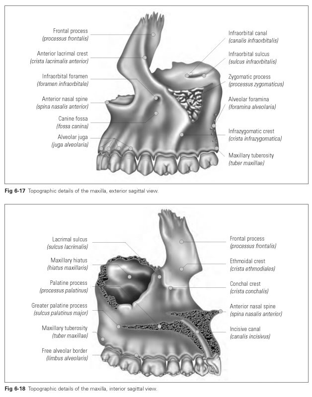
Figures 6-17 and 6-18 illustrate the topographic details of the maxilla.