Temporomandibular Joint
The mandible as a single bone is fixed to the skull by two true (synovial) joints. The two joints are entirely separate from each other, but because they are identical in structure and because movement of the mandible always takes place simultaneously in both joints, they are often merely referred to as a single joint (articulatio temporo-mandibularis; Latin, articulatio = joint). Therefore, the topographic description can be applied to a single TMJ.
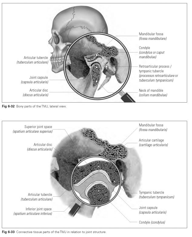
The TMJ is located directly anterior to the opening of the outer auditory canal (porus acusticus externus). A distinction is made between the bony and the connective tissue parts of the joint. The bony parts of theTMJ (Fig 6-32) include the:
- Mandibular fossa (fossa mandibularis)
- Articular tubercle (tuberculum articulare; emi-nentia articularis)
- Retroarticular process (processus retroarticu-lare; or tympanic tubercle: tuberculum tympani-cum); these parts belong to the squamous part of the temporal bone and are the fixed parts of the joint
- Condyle (condylus or caput mandibulae), a part of the mandible and the movable part of the joint
The connective tissue parts of the TMJ (Fig 6-33) include the:
- Joint capsule (capsula articularis)
- Articular disc (discus articularis)
- Articular cartilage (cartilago articularis)
- Articular ligaments (ligamenta)
Form and position of the individual parts of the joint
The articular surface belongs to the squamous part of the temporal bone so that the joint actually permits movement between the temporal bone and the mandible, hence the name temporomandibular joint. The actual mandibular fossa is bounded posteriorly by the root of the zygomatic process of the temporal bone. This part is known as the retroarticular process or tympanic tubercle. The anterior border of the mandibular fossa forms the articular tubercle (tuberculum articulare).
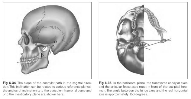
The articular surface is a long depression whose transverse axis meets the transverse axis of the opposing joint at an obtuse angle in front of the occipital foramen. In addition, the articular surface viewed laterally forms an S-shaped curve, where the concave surface from the depth of the mandibular fossa is inclined anteriorly, sloping downward, and becomes a convex section. This surface is known as the condylar path. The inclination of the condylar path in the sagittal plane (Fig 6-34) can be determined as the horizontal condylar inclination in relation to various reference planes (auriculo-infraorbital plane, Frankfort plane, Camper plane).
The articular surface is covered with smooth, elastic fibrocartilage that reduces pressure and friction. In the bottom of the mandibular fossa, the articular surface is made up of only a thin bony plate, which can scarcely be loaded by pressure; this indicates that masticatory pressure is not transferred in the mandibular fossa but to other bony parts of the joint.The articular tubercle and the inner and outer marginal ridges are the most suitable for functional loading.
The condyle (caput mandibulae; condylus) is located on the articular process of the mandible. It resembles a kidney-shaped roll of bone whose transverse axis is adapted to the articular surface (Fig 6-35); ie, the transverse axes of the two condyles do not form a line but intersect at an obtuse angle (approximately 150 to 165 degrees).
The anterior part of the condyles is covered with cartilage and is much smaller than the mandibular fossa. The resulting lack of closeness of fit is known as incongruence.
The articular disc (discus articularis) lies between the mandibular fossa and the condyle and compensates for the incongruence between these parts of the joint. This disc divides the joint into two parts because it is connected to the joint capsule all the way around.
The articular capsule (capsula articularis) is a loose casing of connective tissue that forms an airtight seal around all parts of the joint so that slight negative pressure is created inside the joint and the joint components are pressed together. The external layer of the capsule (membrana fibrosa) is made up of bundles of collagen fibers, while the internal layer (membrana synovialis) carries blood vessels and nerves and secretes the synovial fluid. The joint capsule is fixed around the articular surface on the temporal bone and on the condyle.