Features of the face
The human face (facies) can be divided into three areas by the interpupillary line (a line through the pupils with the eyes looking straight ahead; see Fig 1-10) and a line through the oral aperture:
- Upper face, from the hairline over the forehead (frons) to the eyebrows (supercilium)
- Middle part of the face, including the maxilla, nose, and upper lip
- Lower face, including the mandible, from the orifice of the mouth (rima oris) to the chin (menton)
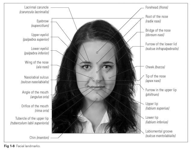
At the sides, the face extends over the temple region (tempus) and the external ear (auricula) as far as the posterior and inferior edge of the mandible. From the frontal view, cephalometric (or anthropometric) measuring points can be established, which are used for analysis of the facial profile.The facial landmarks described in this section are illustrated in Fig 1-8.
The eyeballs can be covered by both eyelids (palpebra superior/inferior), the upper and lower edge of the lids forming the palpebral fissure (rima palpebrarum). In the medial angle of the eye at the root of the nose, there is a small mucosal projection, the lacrimal caruncle (caruncula lacrimalis).
The external part of the nose projects from the midface. The nose extends from its root (radix nasi), which is located between the eyes, to the bridge (dorsum nasi) and finally to the mobile tip (apex nasi), whose sides or wings (alae nasi) form the entrance to the nose or the nasal vestibule.
The orifice of the mouth (rima oris) forms the entrance to the oral cavity, which is enclosed by the upper and lower lips (labium superius and inferius). When the mouth is closed, the orifice forms a curved line.
The angles of the mouth, where the upper and lower lips meet, lie level with the maxillary canine teeth.The upper lip is bordered above by the nose and at the sides by the nasolabial sulcus (sulcus nasolabialis). The philtrum, a flat furrow, divides the upper lip into two halves and forms the tubercle (tuberculum labii superioris).
In the middle of the lower lip, there is a gentle depression into which the tubercle of the upper lip fits. Below this, the labiomental groove (sulcus mentolabialis) delineates the lower lip from the chin area.
The lips (labia oris) are folds of skin that mainly contain muscles but also glands. Three different types of skin are found in the lip area:
- The outer skin section (pars cutanea) up to the nose or the chin. In men, this may be largely covered with hair and well supplied with sebaceous glands.
- The transitional area (pars intermedia), which forms the red lip margin and is responsible for the redness of the lips. There are no sebaceous glands or hair in this area.
- The mucosal area (pars mucosa), the section of the lips up to the oral vestibule that contains the lip glands with outlets into the vestibule.
In the fornix or trough (fornix vestibuli), the lips intersect with the gingiva, which is attached to the jaws. Lip shape and fullness are essentially achieved by the maxillary anterior teeth. The upper lip lies directly over the labial surfaces, while the lower lip gently bulges outward over the incisal edges of the maxillary anterior teeth. Therefore, the natural and functionally ideal position of the maxillary anterior teeth must be carefully reconstructed during dental treatment for each individual patient.
The distance between the mouth and the nose is smaller than that between the mouth and the chin. In many cases, the space between the nose and the mouth is roughly half the distance to the chin. In other cases, the upper lip is even shorter so that the distance from the orifice of the mouth to the chin is three times that to the nose. When a person laughs, the orifice of the mouth widens,
and the angles of the mouth can be drawn back to behind the second premolars, so that the maxillary anterior teeth and gingiva may become visible.
The lip muscles make the mouth area one of the most mobile parts of the body. The lips play an important role in the creation of facial expressions; they take on characteristic shapes during speaking, smiling, frowning, and otherwise nonverbally expressing emotion and are also involved in the ingestion of food.
The cheeks (buccae) originate from the sides of the face at the nasolabial sulcus (sulcus nasola-bialis) and, together with the lips, form the external border of the oral vestibule. The cheeks can contain thick pads of fat; the layer of fatty tissue in the faces of women may be twice as thick as in men.The thickness of facial skin varies across the different sections of the face, but it is highly elastic in all areas and has a plentiful supply of blood vessels and nerves.
The muscular basis of the cheeks is formed by the buccinator muscle (musculus buccinator), which attaches the cheeks to the molars and premolars.The mucosa of the cheeks, like that of the lips, contains small mixed salivary glands (glan-dulae buccales). The exit point of the parotid gland in the form of a small mucosal protuberance (papilla parotidea) is located close to the second molar, and this is the reason for the tartar deposits that are commonly found on the vestibular surface of the maxillary molars.
The chin (menton) is a characteristic feature of the human face that developed as the cranium grew larger and the jaw became shorter. Owing to the development of speech, the tongue became larger, and the arch of the mandible widened. This resulted in a significant buildup of bone at the middle fusion line of the two halves of the mandible (symphysis) in order to absorb initial transverse stresses. After birth, the chin develops as the primary teeth are shed. The chin may be wide, narrow, long, short, square, oval, or round and, depending on the occlusal relationship, may either protrude or recede. It also may be indented to a lesser or greater extent by a dimple.