Maxillary incisors
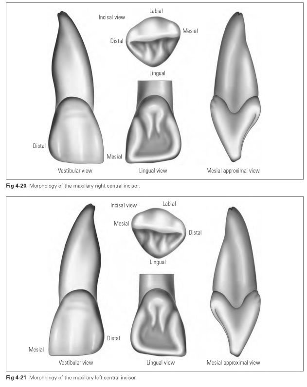
The maxillary central incisor (dens incisivus me-dialis) (Figs 4-19 to 4-21) is the largest incisor tooth, having a rectangular to rhomboid, triangular, or oval vestibular surface, which can harmonize with the shape of the face.
Its average dimensions are:
- Crown width (mesiodistal): 8.5 mm
- Crown depth (labiolingual): 7.0 mm
- Crown length: 11.5 mm
- Total apicocoronal length: 25.0 mm
Its root forms an elongated cone, is straight from an approximal view, and is slightly bent distally from a labial view (root characteristic). The strong root makes it usable as an abutment tooth or suitable for placement of a post crown.
The labial surface (see Fig 4-19a) is almost rectangular and tapers cervically. The mesial edge looks rather straight, whereas the distal edge is curved. The labial surface has a vertical and horizontal curvature characteristic as well as a pronounced angle characteristic. The surface is defined by two marginal ridges and a medial ridge, which are separated by two vertical grooves. When the teeth are freshly erupted, these ridges make the incisal margin appear to have three lobes. This form is lost through abrasion. The distal approximal edge is slightly concave to the cervix and becomes an evenly curved cervical margin. A few slightly pronounced cervical grooves run almost parallel to the cervical margin.
The lingual surface (see Fig 4-19b) resembles the labial surface but is smaller and narrower and tapers more sharply, especially in the cervical area. Two marginal ridges run from the incisal edge cervically, then merge in the apical third of the surface to form the dental tubercle. The marginal ridges outline the concave surface like the edges of a shovel. Differently shaped medial ridges emerge from the tubercle. Depending on the concavity of the lingual surface and the number of enamel ridges, the tubercle can be divided into two tubercles, for example, or the margins may form a partially covered canal.
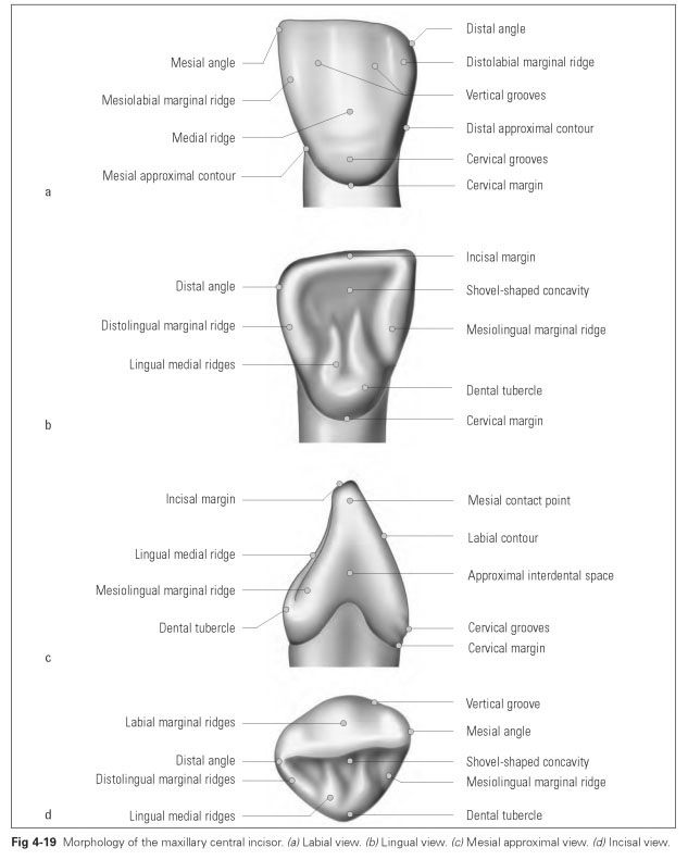
The mesial approximal surface (see Fig 4-19c) has a triangular shape, with the apex at the incisal edge and the baseline at the cervix; the functional chisel shape of the incisor can be seen. The cervical line curves considerably in an incisal direction. The surface itself is tapered toward the neck of the tooth. The vertical curvature of the labial surface contour and the prominent dental tubercle, whose curvature is greatest at the neck, can be clearly seen. The mesial approximal surface is larger than the distal surface.
The incisal view (see Fig 4-19d) shows the features of the other surfaces described above: The curvature characteristic can be seen, and the distal aspect of the surfaces tapers slightly, while the mesial aspect is prominently curved.The cusplike thickening of the dental tubercle can be clearly seen, as can the marginal ridges and the medial ridge. Figure 4-19d shows both the mesial and the distal contact points as the widest convexity of the approximal surfaces.
The maxillary lateral incisor (dens incisivus lateralis) (Figs 4-22 and 4-23) has the same basic shape as the central incisor but is significantly smaller.
Its average dimensions are:
- Crown width (mesiodistal): 6.5 mm
- Crown depth (labiolingual): 6.0 mm
- Crown length: 10.0 mm
- Total apicocoronal length: 23.0 mm
Variations in form and size can be seen with this tooth. It is one of the teeth that is in the process of regression; it may sometimes be entirely absent. It can resemble a primary tooth; even pointed, peg, and pin shapes are possible. The dental tubercle may be developed into a separate lingual tubercle. The lateral incisor is rather shorter than the central incisor, but it is inclined mesially like the central incisor. It has a greater vestibular inclination than the central incisor. The second incisor is single-rooted with a distinctive root characteristic: The root is flattened in the mesiodistal direction and poorly developed with lateral longitudinal fissures, and the
root canal is often deformed.
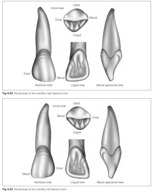
The labial surface is the same as that of the central incisor but smaller and more delicate with rounded edges and more pronounced curvature and angle characteristics.
The lingual surface has very pronounced marginal ridges and a distinctive tubercle.The surface often appears concave and undercut, especially if the tubercle is very strongly developed and the surface is rather covered over. This is where caries often develops.
The approximal surfaces are markedly concave below the contact points, so that the labial surface appears strikingly triangular. Form variations in the lateral incisors are particularly noticeable in this view.
The incisal view shows a very rounded incisal margin, and all the curvatures are clearly visible.