Denture-Bearing Area in the Mandible
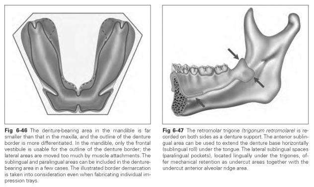
The mandibular denture does not rest on a large bony foundation, unlike the support provided by the palate in the maxilla. Owing to the small area available (only the alveolar ridge with the dorsally positioned trigones), the retaining effect is also considerably reduced.The bony foundation in the mandible is highly variable in its atrophied forms. From a relatively well-developed, high, sharp alveolar ridge to a totally flat alveolar ridge that may even lie inferior to the floor of the mouth, all forms are possible.
After tooth loss, the alveolar ridge of the mandible is also resorbed in the direction of inclination, which gives rise to the particular form of the alveolar line: The straight alveolar lines bend sharply at precisely the canine point; the alveolar line of the posterior teeth widens out, while it sinks down in a lingual direction in the anterior area. A trapezoid open at one side is formed by the parabolic alveolar line.
As well as the relatively small denture-bearing area of the alveolar ridge, the complicated marginal course of the movable buccal mucosa and especially the highly mobile floor of the mouth impede retention of a complete denture. In addition, the shape, size, and mobility of the tongue have an influence on shaping of the denture. A mandibular denture is much more exposed to the muscle activities of the cheeks, lips, tongue, and floor of the mouth than a maxillary denture.
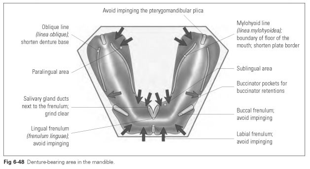
The outward course of the bony wall of the mandibular body means that the vestibular fornix is usually underlaid with bone in the vestibular area. In the lingual region, the sturdy mylohyoid ridge marks the limit of the denture border. As a result, the border of the mandibular denture has very definite areas requiring reduction (Fig 6-46).
The inferior labial frenulum (frenulum labii inferioris) and buccal frenulum (frenulum buccae) should not be impinged, as in the maxilla, so as not to impede movement of the ligaments.
The mental foramen (foramen mentale) may lie on the alveolar ridge if there has been severe atrophy, and it must then be avoided or covered. If there are pressure points in this area, the patient may experience pain from teeth that are no longer there.
The oblique line (linea obliqua), as the insertion of the buccinator muscle, forms the buccal boundary of the base, from which premolars run dorsally. The denture border must be shortened in keeping with this line.
The pterygomandibular plica (plica pterygo-mandibularis or raphe pterygomandibularis; plica = fold; raphe = seam) runs from the posterior border of the retromolar trigone dorsally and upward to the maxillary tuberosity and must not be impinged, as in the maxilla.
The mylohyoid line (linea mylohyoidea), as the insertion of the mylohyoid muscle and the boundary of the floor of the mouth, also demarcates the border of the denture plate. This is because during swallowing the floor of the mouth is raised along this line, and a plate border that extends beyond this line in the floor of the mouth will cause severe pressure points. The patient will often be unable to swallow without difficulty. The edge of the plate, from the premolars onward, should therefore be shortened dorsally as far as the retromolar trigones in keeping with this line.
The frenulum of the tongue (frenulum linguae) must not be impinged because, if impinged, the mobility of the tongue will be impeded. If the denture base is to be reground, only the insertion of the frenulum needs to be avoided. The denture border should not be widened along the course of the frenulum because that will impede the suction capacity of the denture base.
The sublingual caruncles (carunculae sublingualis) are small papillae on the left and right next to the frenulum of the tongue and are openings of the salivary glands. An impression must not be taken of these openings because they can become blocked, preventing the flow of saliva. The result for the patient is a dry mouth, with the salivary gland swelling up and causing an unpleasant feeling of tightness. The impressions of the papillae can be created in the denture border by grinding out slightly.
The alveolar tubercle of the mandible (tubercu-lum alveolare mandibulae) is a bulge of mucous membrane over the retromolar trigone (trigonum retromolare) that can arise because of particular muscle attachments. The denture base can be extended as far as this (Fig 6-47), but the pterygomandibular plica must not be impinged.
The alveolar jugae (juga alveolaria) are prominences over the roots of the teeth, which should only be reproduced in the mandible in the area of the anterior teeth, whereas the dorsal area of the
outer edge of the denture should be kept smooth for hygiene reasons. Any manipulation of the denture border should be done by the dentist and not by the dental technician.
Figure 6-48 illustrates the denture-bearing area in the mandible.