Muscle Groups of the Hyoid Bone
Several functional groups of muscles attach to the hyoid; these are involved in both jaw movement and the swallowing process. According to the theoretical classification, they can be characterized as mouth-opening muscles. A distinction is made between the suprahyoid muscles (above the hyoid) and the infrahyoid muscles (below the hyoid).
Suprahyoid muscles (musculi suprahyoidei) form a functional group of muscles that are all fixed above the hyoid bone (Figs 7-41 and 7-42). They form the floor of the mouth and are not part of the actual masticatory musculature but act as mouth-opening muscles. During swallowing, they pull the hyoid with the larynx up and forward and raise the floor of the mouth and tongue. In the process, the mandible is fixed in terminal occlusion by the masticatory muscles. The following paragraphs outline these muscles in detail.
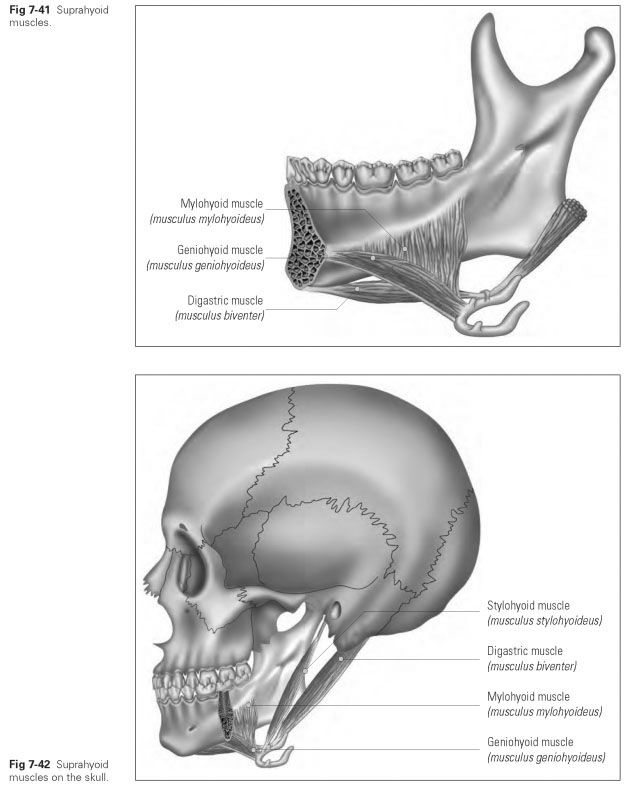
The digastric muscle (musculus biventer or musculus digastricus) has its origin at the inferior edge of the mandible in the digastric fossa. This first belly of the muscle (venter anterior) lies under the floor of the mouth and joins at the hyoid to form a round intermediate tendon, which is able to slide back and forth here in a fibrous loop. From there, the second belly (venter posterior) runs at an obtuse angle downward and upward to the digastric sulcus of the mastoid process on the temporal bone (sulcus digastricus processus mastoideus). When both bellies of the muscle contract, the hyoid is moved upward and forward. With the hyoid fixed, the mandible is opened.The bellies of the muscle are able to contract separately from each other.
The mylohyoid muscle (musculus mylohyoi-deus) is known as the muscle of the floor of the mouth and, together with the muscle on the opposite side and the connecting fused tendon, as the diaphragma oris. This paired muscle originates at the mylohyoid line on the interior of the mandible. This line of origin extends dorsally to the anterior edge of the medial pterygoid muscle. The mylohyoid muscle attaches in the middle of the hyoid body and, with the muscle bundle on the opposite side, forms a raphe that extends from the middle of the hyoid to the symphysis of the mandible (raphe mylohyoidea). When this muscle is in its resting position, the hyoid lies roughly level with the mandibular angle. However, the mylohyoid line lies much higher dorsally so that posteriorly both muscle bundles are at sharp angles to each other. On contraction, they pull the floor of the mouth and the hyoid upward to support swallowing, because raising the floor of the mouth presses the tongue against the palate.
The geniohyoid muscle (musculus geniohyoi-deus) originates at the inferior mental spines on the interior of the mandible (spina mentalis mandibulae) and, as a thick muscle bundle, runs to the superior edge of the body of the hyoid (corpus ossis hyoidei). When the hyoid is fixed in its position by the middle constrictor muscle of the pharynx and the infrahyoid muscles, the geniohyoid muscle is able to draw the mandible back. In centric occlusion, this muscle pulls the hyoid upward and forward, which supports the swallowing process. The geniohyoid muscle lies on the mylohyoid muscle.
The stylohyoid muscle (musculus stylohyoi-deus) originates on the dorsal side of the styloid process of the temporal bone (processus styloi-deus), divides into two bellies, and attaches to the body of the hyoid and the greater horn of the hyoid; it encloses the intermediate tendon of the digastric muscle. It draws the hyoid upward and backward.
The infrahyoid muscles (musculi infrahyoidei) form a functional group of muscles that are all fixed below the hyoid bone.They fix the hyoid and prevent it from rising when the mouth is opened. There are four muscles in this muscle group:
- The sternohyoid muscle (musculus sternohyoi-deus) has its origin on the sternum and its insertion on the body of the hyoid.
- The sternothyroid muscle (musculus sternothy-roideus) has its origin on the sternum and its insertion on the oblique line of the thyroid cartilage.
- The thyrohyoid muscle (musculus thyreohyoi-deus) runs from the oblique line of the thyroid cartilage to the body and greater horn of the hyoid.
- The omohyoid muscle (musculus omohyoideus) has its origin at the superior border of the clavicle and its insertion on the body of the hyoid.